. 2016 Mar;8(3):600-7.
doi: 10.21037/jtd.2016.01.60.

Video-assisted thoracoscopic surgery resection and reconstruction of thoracic trachea in the management of a tracheal neoplasm

Affiliations

Video-assisted thoracoscopic surgery resection and reconstruction of thoracic trachea in the management of a tracheal neoplasm

Shuben Li et al. J Thorac Dis. 2016 Mar.

Abstract

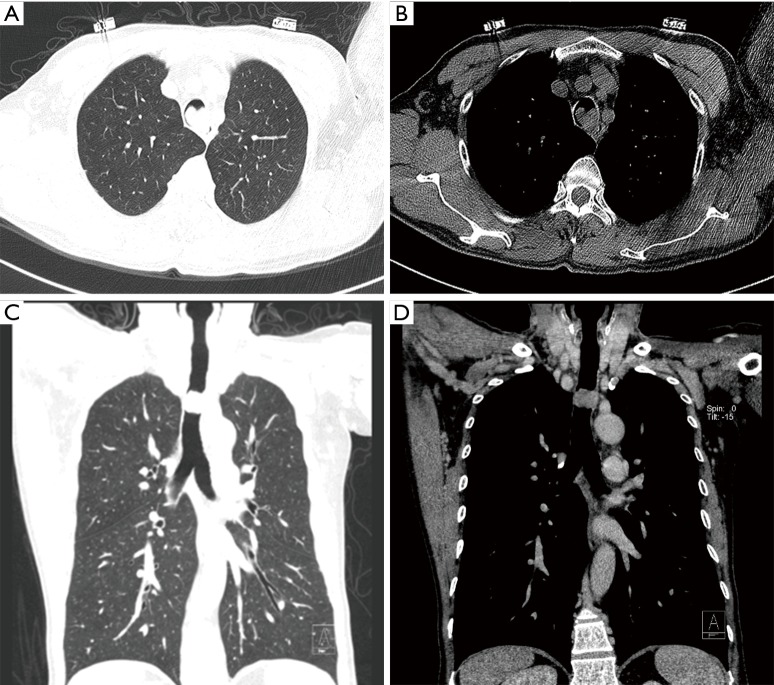
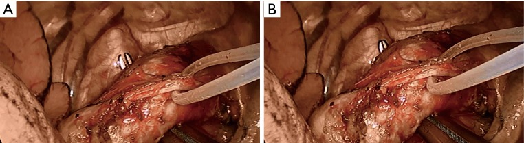
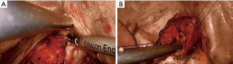
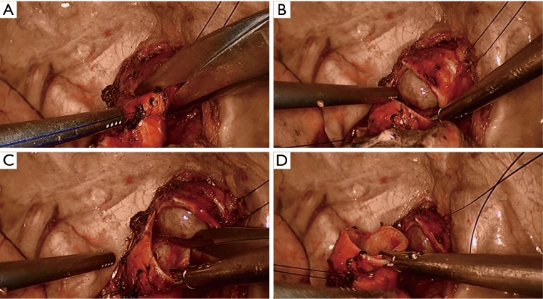
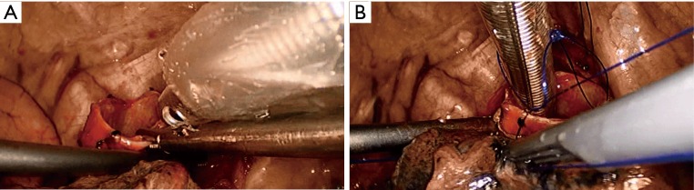
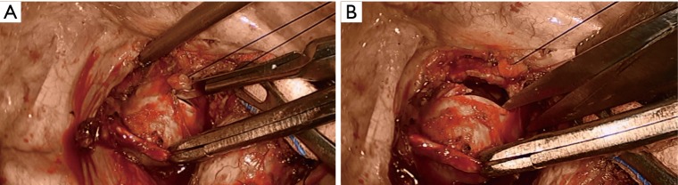
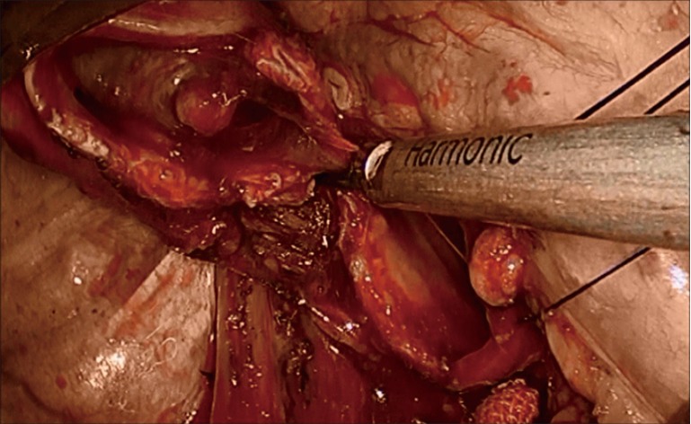
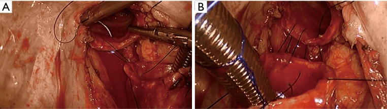
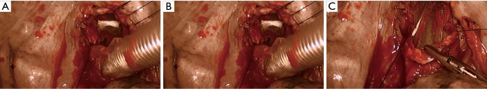
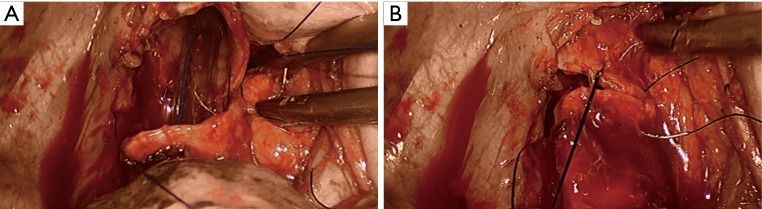
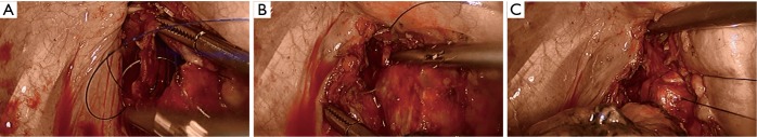
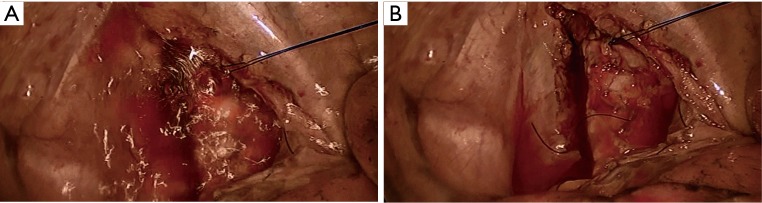
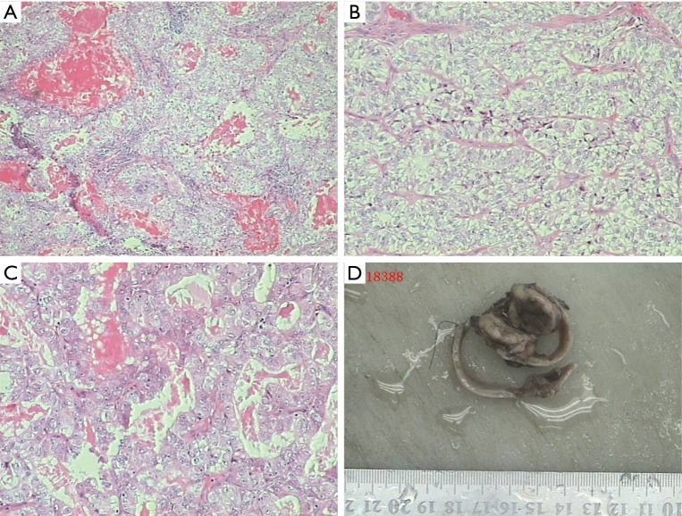
Intratracheal tumor is a rare tumor, accounting for only 2% of upper respiratory tract neoplasms. Its symptoms are similar to those of head and neck cancers, including coughing up blood, sore throat, and airway obstruction. The diagnosis of this disease is often based on the findings of fibrobronchoscopy or computed tomography (CT). Surgery remains the treatment of choice for tracheal tumor. In patients with benign neoplasms or if the tumors have limited involvement, fibrobronchoscopic resection of the tumor can be performed. For malignant tumors, however, radical resection is required. In the past, open incision is used during the surgery for tumors located in thoracic trachea. Along with advances in video-assisted thoracoscopic surgery (VATS) minimally invasive techniques and devices, VATS resection and reconstruction of the trachea can achieve the radical resection of the tumor and meanwhile dramatically reduce the injury to the patients. In this article we describe the application of VATS resection and reconstruction of trachea in the management of a tracheal neoplasm.

Keywords: Tracheal tumor; resection; surgery; thoracoscopy; video-assisted; video-assisted thoracoscopic surgery (VATS).


Conflict of interest statement

Conflicts of Interest: The authors have no conflicts of interest to declare.

Figures

Figure 1
Figure 1
The preoperative computed tomography (CT) showed a mass inside the middle portion of the thoracic trachea; the tracheal lumen where the mass was located became obviously narrow, and the mass blocked most of the tracheal lumen.
Figure 2
Figure 2
Open the mediastinal pleura to dissociate thoracic trachea.
Figure 3
Figure 3
Disassociate the tissues behind the thoracic trachea.
Figure 4
Figure 4
Thoroughly dissociate the trachea and then suspend it using a suture.
Figure 5
Figure 5
Cut the trachea at distal end of the tumor using tissue scissors. It was found that the roundish tumor had smooth surface and was latticed with tiny blood vessels; it was not obviously lobulated, showing a translucent status.
Figure 6
Figure 6
The tracheal tube was placed at the distal stump via the thoracic cavity and then fixed with sutures.
Figure 7
Figure 7
The trachea was cut open at the proximal end of the tumor using a scalpel, and the proximal stump was pruned using tissue scissors.
Figure 8
Figure 8
The posterior wall of the trachea was divided using HIFU to achieve the en bloc resection of the tumor and the involved part of the trachea.
Figure 9
Figure 9
The posterior wall of the trachea was continuously closed using the 2–0 Prolene sutures.
Figure 10
Figure 10
The tracheal tube was withdrawn via the thoracic cavity and then inserted via mouth.
Figure 11
Figure 11
The anterior wall of the trachea was continuously closed using the 2–0 Prolene sutures.
Figure 12
Figure 12
The lateral wall of the trachea was continuously closed using the 2–0 Prolene sutures.
Figure 13
Figure 13
Anastomotic leak testing; knots were tied and fixed after the anastomosis was confirmed to be leak-free.
Figure 14
Figure 14
Pathology: the tumor tissue was adhered to the tracheal wall and had relatively clear boundary with the wall. The tumor cells showed acinar-like architecture, with clear cytoplasm. The cytologic atypia was not obvious, and no mitotic figure was observed. Within the acinar lumen there were a large number of mucus-like secretions. The lesion was confirmed to be low-grade malignant adenocarcinoma that might have been arisen from tracheal glands (magnification, ×100).
Figure 15
Figure 15
Video-assisted thoracoscopic surgery (VATS) resection and reconstruction of thoracic trachea (1). Available online: http://www.asvide.com/articles/912

Comment in

References

    1. Li S, Liu J, He J, et al. Video-assisted thoracoscopic surgery (VATS) resection and reconstruction of thoracic trachea. Asvide 2016;3:157. Available online: http://www.asvide.com/articles/912 - PMC - PubMed
    1. Bhattacharyya N. Contemporary staging and prognosis for primary tracheal malignancies: a population-based analysis. Otolaryngol Head Neck Surg 2004;131:639-42. 10.1016/j.otohns.2004.05.018 - DOI - PubMed
    1. Grillo HC. Development of tracheal surgery: a historical review. Part 1: Techniques of tracheal surgery. Ann Thorac Surg 2003;75:610-9. 10.1016/S0003-4975(02)04108-5 - DOI - PubMed
    1. Licht PB, Friis S, Pettersson G. Tracheal cancer in Denmark: a nationwide study. Eur J Cardiothorac Surg 2001;19:339-45. 10.1016/S1010-7940(01)00597-8 - DOI - PubMed
    1. Schraube P, Latz D, Wannenmacher M. Treatment of primary squamous cell carcinoma of the trachea: the role of radiation therapy. Radiother Oncol 1994;33:254-8. 10.1016/0167-8140(94)90360-3 - DOI - PubMed

LinkOut - more resources