Review
. 2015 May 20:4:10-6.
doi: 10.1016/j.bdq.2015.04.002. eCollection 2015 Jun.

Proximity assays for sensitive quantification of proteins

Affiliations
Review

Proximity assays for sensitive quantification of proteins

Christina Greenwood et al. Biomol Detect Quantif. .

Abstract

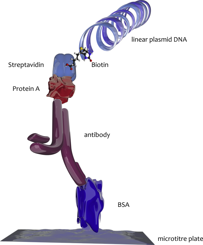
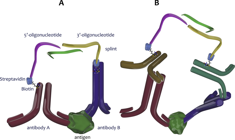
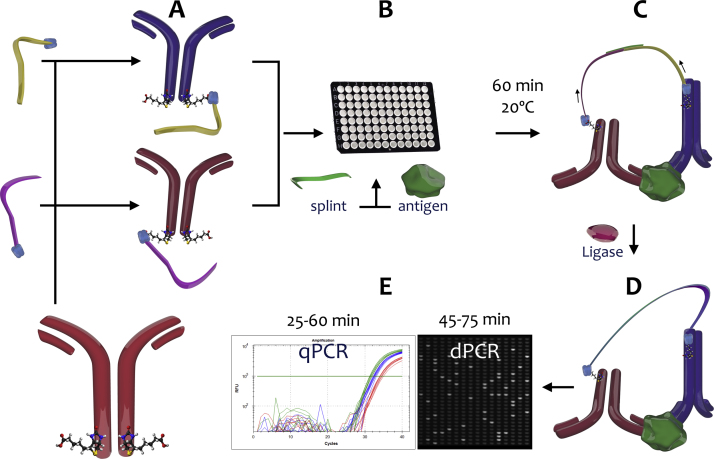
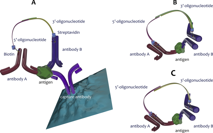
Proximity assays are immunohistochemical tools that utilise two or more DNA-tagged aptamers or antibodies binding in close proximity to the same protein or protein complex. Amplification by PCR or isothermal methods and hybridisation of a labelled probe to its DNA target generates a signal that enables sensitive and robust detection of proteins, protein modifications or protein-protein interactions. Assays can be carried out in homogeneous or solid phase formats and in situ assays can visualise single protein molecules or complexes with high spatial accuracy. These properties highlight the potential of proximity assays in research, diagnostic, pharmacological and many other applications that require sensitive, specific and accurate assessments of protein expression.

Keywords: Immuno-PCR; Immunoassays; In situ assays; Proximity extension assay; Proximity ligation assay.


Figures

Fig. 1
Fig. 1
The original iPCR made use of a recombinant streptavidin-protein A chimera with bispecific affinity for DNA and antibodies to link linear plasmid DNA to an antibody specific for bovine serum albumin (BSA), which was immobilised on the surface of microtitre wells. Binding of the antibody to BSA resulted in a specific antigen–antibody–DNA conjugate that was detected by agarose gel electrophoresis after PCR amplification with plasmid-specific primers.
Fig. 2
Fig. 2
Direct and indirect PLA. (A) Biotinylated antibodies bind pairwise to adjacent epitopes on target proteins. This brings the two streptavidin-oligonucleotide tails, one coupled through its 5′-end, the other through its 3′-end, into close proximity. The connector oligonucleotide (splint) hybridises to both oligonucleotides, resulting in adjacent free 3′-OH and 5′-phosphate moieties. The gap is ligated and the resulting continuous DNA strand can be amplified and detected. (B) The indirect form of PLA follows the same principle, except that unmodified primary antibodies are detected with secondary antibodies that are conjugated to the DNA strands.
Fig. 3
Fig. 3
PLA workflow. (A) Oligonucleotides synthesised with a streptavidin molecule at their 5′- or 3′-ends are combined with biotinylated antibodies to form proximity probes. (B) Probes, sample and splint are combined and both probes bind simultaneously to their epitopes on the target antigen, if present, and the 5′- and 3′-oligonucleotides are joined by the splint. (C) DNA ligase connects the gap and thus joins the two oligonucleotides. (D) This generates a full length DNA amplicon that can be amplified and detected by several methods. (E) Detection by qPCR or dPCR is shown.
Fig. 4
Fig. 4
A. Solid phase PLA. (A) Capture antibody immobilised in a microtitre well binds target antigen, with unbound particles and other sample components removed by washes. Proximity probes are then added to the well and a PLA is carried out. (B) First generation PEA. The 3′-oligonucleotide is double stranded with a 3′-overhang. In the presence of target antigen, the probe oligonucleotides can hybridise to each other, leading to the extension of one oligonucleotide into a DNA template that can be detected and quantified. (C) Second generation PEA. Single stranded oligonucleotides hybridise directly to each other, with one becoming extended by a DNA polymerase to generate an amplifiable DNA template.

Similar articles

Cited by

References

    1. Bustin S.A. IUL Press; La Jolla, CA: 2004. A–Z of quantitative PCR.
    1. Higuchi R., Fockler C., Dollinger G., Watson R. Kinetic PCR analysis: real-time monitoring of DNA amplification reactions. Biotechnology (NY) 1993;11:1026–1030. - PubMed
    1. Gibson U.E., Heid C.A., Williams P.M. A novel method for real time quantitative RT-PCR. Genome Res. 1996;6:995–1001. - PubMed
    1. Heid C.A., Stevens J., Livak K.J., Williams P.M. Real time quantitative PCR. Genome Res. 1996;6:986–994. - PubMed
    1. Engvall E., Perlmann P. Enzyme-linked immunosorbent assay (ELISA). Quantitative assay of immunoglobulin G. Immunochemistry. 1971;8:871–874. - PubMed