

PET/CT imaging of Mycobacterium tuberculosis infection
- PMID: 27077068
- PMCID: PMC4820496
- DOI: 10.1007/s40336-016-0164-0
PET/CT imaging of Mycobacterium tuberculosis infection
Abstract
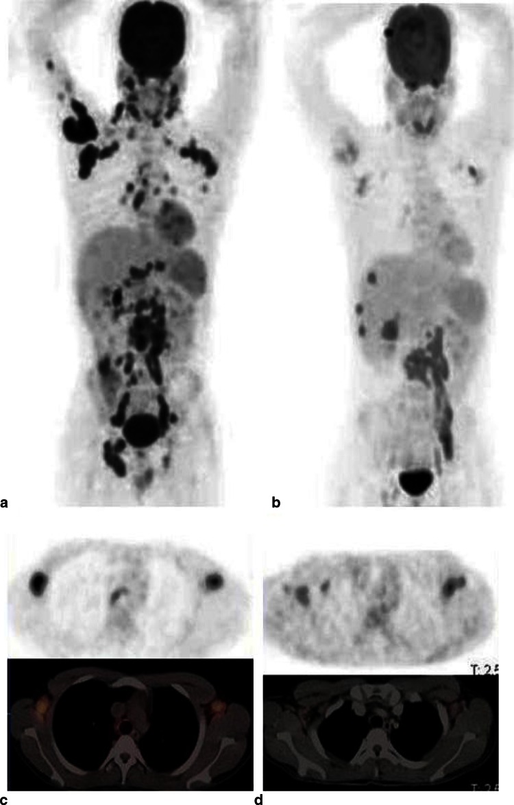
Tuberculosis has a high morbidity and mortality worldwide. Mycobacterium tuberculosis (Mtb) has a complex pathophysiology; it is an aerobic bacillus capable of surviving in anaerobic conditions in a latent state for a very long time before reactivation to active disease. In the latent tuberculosis infection, the individual has no clinical evidence of active disease, but exhibits a hypersensitive response to proteins of Mtb. Only some 5-10 % of latently infected individuals appear to have reactivation of tuberculosis at any one time point after infection, and neither imaging nor immune tests have been shown to predict tuberculosis reactivation reliably. The complex pathology of the organism provides multiple molecular targets for imaging the infection and targeting therapy. Positron emission tomography (PET) integrated with computer tomography (CT) provides a unique opportunity to noninvasively image the whole body for diagnosing, staging and assessing therapy response in many infectious and inflammatory diseases. PET/CT is a powerful noninvasive tool that can rapidly provide three-dimensional views of disease deep within the body and conduct longitudinal assessment over time in one particular patient. Some PET tracers, such as 18F-fluorodeoxyglucose (18F-FDG), have been found to be useful in various infectious diseases for detection, assessing disease activity, staging and monitoring response to therapy. This tracer has also been used for imaging tuberculosis. 18F-FDG PET relies on the glucose uptake of inflammatory cells as a result of the respiratory burst that occurs with infection. Other PET tracers have also been used to image different aspects of the pathology or microbiology of Mtb. The synthesis of the complex cell membrane of the bacilli for example can be imaged with 11C-choline or 18F-fluoroethylcholine PET/CT while the uptake of amino acids during cell growth can be imaged by 3'-deoxy-3'-[18F]fluoro-l-thymidine. PET/CT provides a noninvasive and sensitive method of assessing histopathological information on different aspects of tuberculosis and is already playing a role in the management of tuberculosis. As our understanding of the pathophysiology of tuberculosis increases, the role of PET/CT in the management of this disease would become more important. In this review, we highlight the various tracers that have been used in tuberculosis and explain the underlying mechanisms for their use.
Keywords: 18F-fluoroethylcholine; 3′-Deoxy-3′-[18F]fluoro-l-thymidine; 68Ga-citrate; PET/CT; Tuberculosis.
Figures
References
-
- World Health Organization (2015) Global tuberculosis report 2015 (20th edition): www.who.int/tb/publications/global_report. Assessed 23 Nov 2015
-
- Hershkovitz I, Donoghue HD, Minnikin DE, Besra GS, Lee OY, Gernaey AM, et al. Detection and molecular characterization of 9,000-year old Mycobacterium tuberculosis from a Neolithic settlement in the Eastern Mediterranean. PLoS ONE. 2008;3(10):e3426. doi: 10.1371/journal.pone.0003426. - DOI - PMC - PubMed
Publication types
LinkOut - more resources
Full Text Sources
Other Literature Sources
Research Materials