Review
. 2016 Feb 13:5:30-9.
doi: 10.1016/j.ebiom.2016.02.023. eCollection 2016 Mar.

Modeling Barrier Tissues In Vitro: Methods, Achievements, and Challenges

Affiliations
Review

Modeling Barrier Tissues In Vitro: Methods, Achievements, and Challenges

Courtney M Sakolish et al. EBioMedicine. .

Abstract

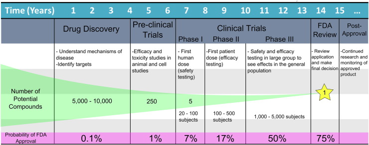
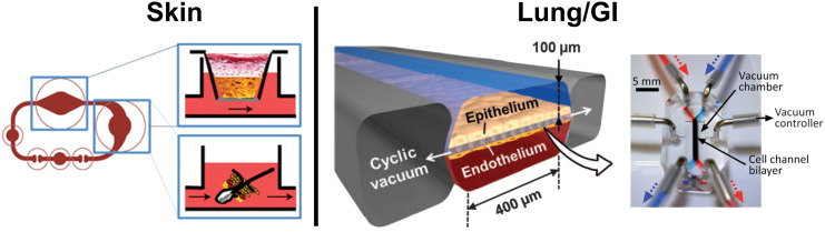
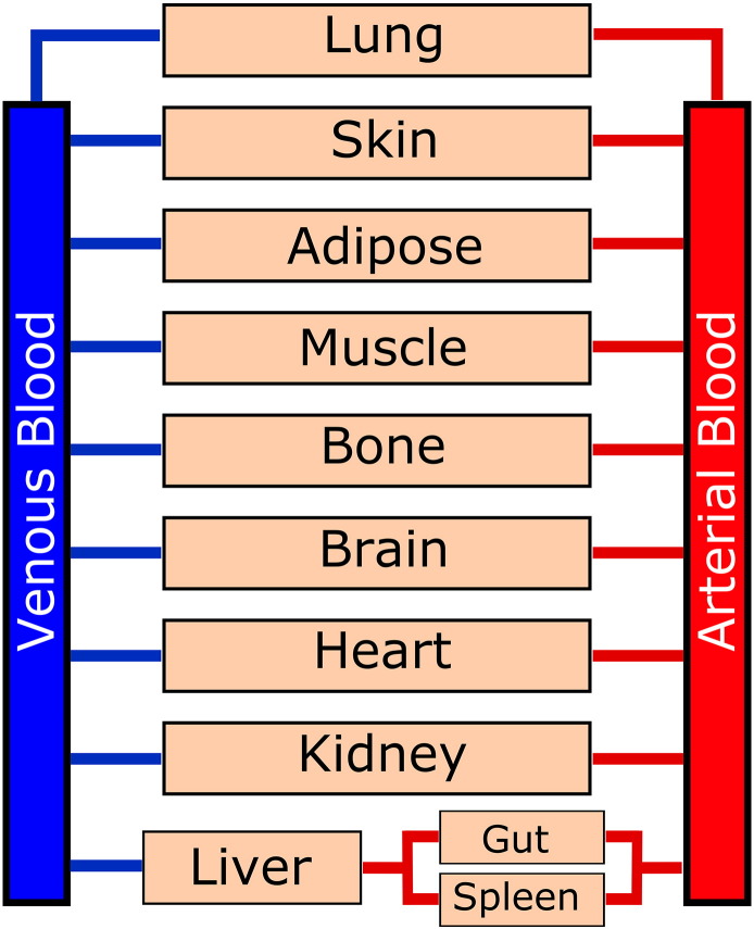
Organ-on-a-chip devices have gained attention in the field of in vitro modeling due to their superior ability in recapitulating tissue environments compared to traditional multiwell methods. These constructed growth environments support tissue differentiation and mimic tissue-tissue, tissue-liquid, and tissue-air interfaces in a variety of conditions. By closely simulating the in vivo biochemical and biomechanical environment, it is possible to study human physiology in an organ-specific context and create more accurate models of healthy and diseased tissues, allowing for observations in disease progression and treatment. These chip devices have the ability to help direct, and perhaps in the distant future even replace animal-based drug efficacy and toxicity studies, which have questionable relevance to human physiology. Here, we review recent developments in the in vitro modeling of barrier tissue interfaces with a focus on the use of novel and complex microfluidic device platforms.

Keywords: Barrier tissues; Drug discovery; In vitro modeling; Microfluidic technologies; Microphysiological systems; Organ-on-a-chip.


Figures

Fig. 1
Fig. 1
Current trends in pharmaceutical development. A model highlighting the time investment, number of potential compounds and probability of approval at each stage of pharmaceutical development from the discovery stage to post-approval (based on information from 2015 CMR International Pharmaceutical R&D Executive Summary and the IFPMA Facts and Figures 2014 report).
Fig. 2
Fig. 2
Mimicking barrier tissues of organs in microdevices. SKIN CHIP: A series of microfluidic chambers were connected to model the skin and hair follicles. A Transwell chamber contained skin biopsies placed over ex vivo subcutaneous tissue with a fluidic basolateral chamber. A fluidic chamber containing follicular hair extracts (FUEs) followed the skin chamber in circuit. Reproduced from Atac et al. (2013) with permission of The Royal Society of Chemistry. LUNG/GI CHIP: This chip design was used to model both the lung and GI barrier tissues. Two fluidic channels run in parallel with a porous membrane to separate epithelium from endothelium. Additionally, empty chambers run along the sides of this device and pull a cyclic vacuum to mimic movement due to breathing or digestion respectively in lung or GI models. GI model reproduced from Kim et al. (2012) with permission of The Royal Society of Chemistry. Lung device from [Huh, D. et al. A human disease model of drug toxicity-induced pulmonary edema in a lung-on-a-chip microdevice. Sci Transl Med, 4, 159ra147]. Reprinted with permission from AAAS.
Fig. 3
Fig. 3
Mimicking barrier tissues of organs in microdevices. KIDNEY CHIP: Kidney tubular epithelial cells were seeded onto an ECM-treated membrane in a fluidic, top channel. Underneath the membrane was a media reservoir to provide nutrients to the basolateral surface of the cell monolayer. Reproduced from Jang and Suh (2010) with permission of The Royal Society of Chemistry. ENDOTHELIUM CHIP: Fully enclosed, perfusable vessels formed in a collagen hydrogel. This hydrogel allows for endothelial growth on all internal surfaces of the vessel, allowing for a close recapitulation of blood vessel formation. Adapted by permission from Macmillan Publishers Ltd.: [NATURE PROTOCOLS] (Morgan et al., 2013), copyright (2013). BLOOD–BRAIN CHIP: A multi-layered microfluidic device with incorporated electrodes for internal TEER measurements. Astrocytes and brain endothelial cells were seeded on opposite sides of the membrane in a back-to-back co-culture resulting in higher resistance to permeability and barrier function than endothelial monocultures. Reproduced from Booth and Kim (2012) with permission of The Royal Society of Chemistry.
Fig. 4
Fig. 4
Physiologically-based pharmacokinetic (PBPK) model of a human system. This model shows the blood circulation throughout the human body through various tissues. Future multi-organ microfluidics will likely be designed following this type of schematic, with various residence times and permeability in each organ chamber.

References

    1. Abaci H.E., Gledhill K., Guo Z., Christiano A.M., Shuler M.L. Pumpless microfluidic platform for drug testing on human skin equivalents. Lab Chip. 2015;15:882–888. - PMC - PubMed
    1. Achyuta A.K., Conway A.J., Crouse R.B., Bannister E.C., Lee R.N., Katnik C.P., Behensky A.A., Cuevas J., Sundaram S.S. A modular approach to create a neurovascular unit-on-a-chip. Lab Chip. 2013;13:542–553. - PubMed
    1. Astashkina A., Mann B., Grainger D.W. A critical evaluation of in vitro cell culture models for high-throughput drug screening and toxicity. Pharmacol. Ther. 2012;134:82–106. - PubMed
    1. Atac B., Wagner I., Horland R., Lauster R., Marx U., Tonevitsky A.G., Azar R.P., Lindner G. Skin and hair on-a-chip: in vitro skin models versus ex vivo tissue maintenance with dynamic perfusion. Lab Chip. 2013;13:3555–3561. - PubMed
    1. Bellin M., Marchetto M.C., Gage F.H., Mummery C.L. Induced pluripotent stem cells: the new patient? Nat. Rev. Mol. Cell Biol. 2012;13:713–726. - PubMed

Publication types