. 2016 Apr 14;11(4):e0153886.
doi: 10.1371/journal.pone.0153886. eCollection 2016.

Accelerated Lactate Dehydrogenase Activity Potentiates Osteoclastogenesis via NFATc1 Signaling

Affiliations

Accelerated Lactate Dehydrogenase Activity Potentiates Osteoclastogenesis via NFATc1 Signaling

Heejin Ahn et al. PLoS One. .

Abstract

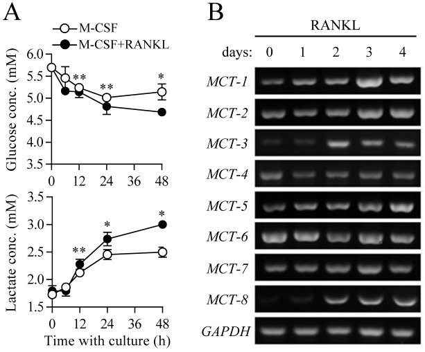
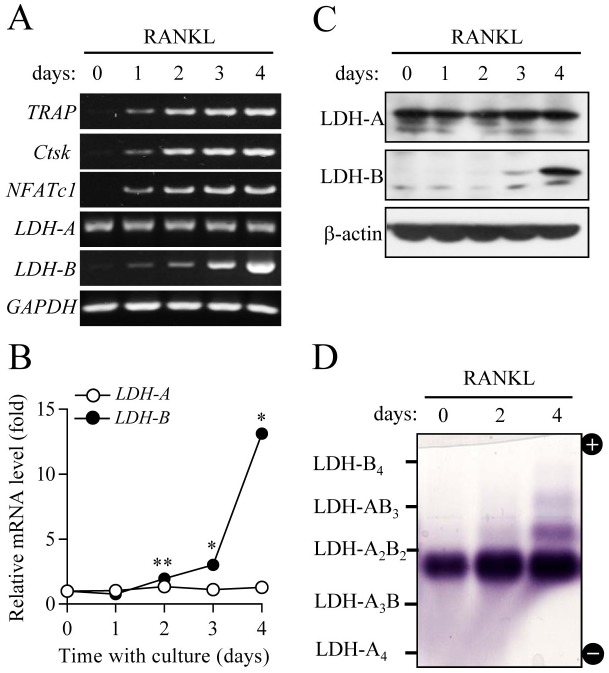
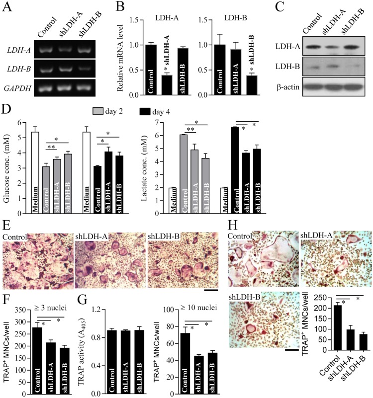
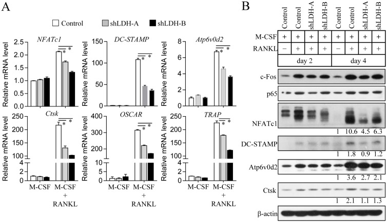
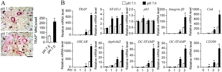
Osteoclasts seem to be metabolic active during their differentiation and bone-resorptive activation. However, the functional role of lactate dehydrogenase (LDH), a tetrameric enzyme consisting of an A and/or B subunit that catalyzes interconversion of pyruvate to lactate, in RANKL-induced osteoclast differentiation is not known. In this study, RANKL treatment induced gradual gene expression and activation of the LDH A2B2 isotype during osteoclast differentiation as well as the LDH A1B3 and B4 isotypes during osteoclast maturation after pre-osteoclast formation. Glucose consumption and lactate production in growth media were accelerated during osteoclast differentiation, together with enhanced expression of H+-lactate co-transporter and increased extracellular acidification, demonstrating that glycolytic metabolism was stimulated during differentiation. Further, oxygen consumption via mitochondria was stimulated during osteoclast differentiation. On the contrary, depletion of LDH-A or LDH-B subunit suppressed both glycolytic and mitochondrial metabolism, resulting in reduced mature osteoclast formation via decreased osteoclast precursor fusion and down-regulation of the osteoclastogenic critical transcription factor NFATc1 and its target genes. Collectively, our findings suggest that RANKL-induced LDH activation stimulates glycolytic and mitochondrial respiratory metabolism, facilitating mature osteoclast formation via osteoclast precursor fusion and NFATc1 signaling.


Conflict of interest statement

Competing Interests: The authors have declared that no competing interests exist.

Figures

Fig 1
Fig 1. Increased glycolytic metabolism during osteoclast differentiation.
Osteoclast precursors were cultured with M-CSF (30 ng/ml) and RANKL (100 ng/ml) for the indicated times. (A) Glucose and lactate contents in cell culture media. Concentrations of glucose and lactate in the culture medium were measured. Data are given as mean ± SD for a representative experiment run in triplicate. *P < 0.01, **P < 0.05. (B) Gene expression analysis of monocarboxylate transporters (MCTs) during osteoclast differentiation. Total RNA isolated from cells was subjected to RT-PCR analysis of the indicated mRNAs. Level of glyceraldehyde 3-phosphate dehydrogenase (GAPDH) served as an internal control for equal loading.
Fig 2
Fig 2. Changes in LDH gene expression and isotypes during osteoclast differentiation.
Osteoclast precursors were cultured in the presence of M-CSF (30 ng/ml) and RANKL (100 ng/ml) for 4 days. (A and B) mRNA levels of LDH-A and LDH-B were determined using RT-PCR (A) and quantitative real-time PCR (B). Data for (B) are means ± SD for a representative experiment run in triplicate. *P < 0.01, **P < 0.05. (C) Protein levels of LDH-A and LDH-B were analyzed using immunoblot analysis. GAPDH and β-actin were used as loading controls. (D) Profiling of LDH isotypes. To analyze LDH isotypes, agarose gel electrophoresis was performed, and the activities were visualized by a formazan color reaction.
Fig 3
Fig 3. Decreased osteoclast formation by LDH-A or LDH-B depletion.
Osteoclast precursors were infected with shRNA lentiviral particles targeting mouse LDH-A, LDH-B, or pLKO.1-puro empty control virus particles. After puromycin selection for 2 days, LDH-A or LDH-B knockdown was validated using RT-PCR (A), quantitative real-time PCR (B), and immunoblot analysis (C). GAPDH and β-actin were used as loading controls. (D) Glucose and lactate concentrations in culture media of LDH-A or LDH-B-depleted cells. LDH-A or LDH-B knockdown cells were cultured with M-CSF (30 ng/ml) and RANKL (100 ng/ml) for 2 or 4 days as indicated. Concentrations of glucose and lactate in the culture media were determined. (E and F) Measurement of osteoclast formation. LDH-A or LDH-B-depleted cells were cultured for 4 days in the presence of M-CSF and RANKL to induce osteoclast formation. Cells were stained with TRAP and photographed using a light microscope (E). Scale bar, 100 μm. The number of TRAP+ MNCs with more than three nuclei was counted under a light microscope (F). (G) Determination of TRAP activity and mature osteoclast formation. To assess the extent of pre-osteoclast formation, TRAP activity was measured on day 2 after osteoclast differentiation (left panel). Mature osteoclast formation was determined by counting the number of TRAP+ MNCs with more than 10 nuclei (right panel). (H) Cell fusion assay. Osteoclast precursors treated with M-CSF and RANKL for 2 days were seeded and further cultured in the presence of M-CSF and RANKL for 3 days. After TRAP staining, TRAP+ MNCs with more than three nuclei were counted using a light microscope. Data are mean ± SD (n = 3). *P < 0.01, **P < 0.05.
Fig 4
Fig 4. Altered osteoclastogenic signaling during osteoclast differentiation of LDH-A or LDH-B-deficient cells.
Osteoclast precursors were infected with LDH-A, LDH-B, or pLKO.1-puro empty control virus particles. LDH-A or LDH-B knockdown cells were cultured with M-CSF (30 ng/ml) and RANKL (100 ng/ml) for 3 days (A) or the indicated days (B). (A) The mRNA expression levels of osteoclastogenic genes including NFATc1, DC-STAMP, Atp6v0d2, cathepsin K (Ctsk), OSCAR, and TRAP were analyzed using quantitative real-time PCR. Data are mean ± SD (n = 3). *P < 0.01. (B) Total cell lysates were subjected to immunoblot analysis with specific antibodies to c-Fos, p65, NFATc1, DC-STAMP, Atp6v0d2, cathepsin K (Ctsk), and β-actin (loading control). Band intensities were represented as a fold difference. Gel images are representative of three independent experiments.
Fig 5
Fig 5. Stimulatory effect of acidic extracellular pH on osteoclast differentiation.
Osteoclast precursors were cultured under a CO2-free condition in HEPES-buffered medium (pH 7.0 or 7.5) in the presence of M-CSF (30 ng/ml) and RANKL (100 ng/ml) for 4 days (A) or for the indicated times (B). (A) Measurement of osteoclast formation. Cells were stained with TRAP and the number of TRAP+ MNCs with more than three nuclei was counted under a light microscope. Scale bar, 100 μm. (B) Analysis of osteoclastogenic gene expression. mRNA levels were analyzed using quantitative real-time PCR.

References

    1. Rodan GA, Martin TJ. Therapeutic approaches to bone diseases. Science. 2000;289(5484):1508–14. . - PubMed
    1. Vignery A. Osteoclasts and giant cells: macrophage-macrophage fusion mechanism. International journal of experimental pathology. 2000;81(5):291–304. - PMC - PubMed
    1. Palokangas H, Mulari M, Vaananen HK. Endocytic pathway from the basal plasma membrane to the ruffled border membrane in bone-resorbing osteoclasts. Journal of cell science. 1997;110 (Pt 15):1767–80. . - PubMed
    1. Teitelbaum SL, Ross FP. Genetic regulation of osteoclast development and function. Nature reviews Genetics. 2003;4(8):638–49. 10.1038/nrg1122 . - DOI - PubMed
    1. Boyle WJ, Simonet WS, Lacey DL. Osteoclast differentiation and activation. Nature. 2003;423(6937):337–42. 10.1038/nature01658 . - DOI - PubMed

Publication types

MeSH terms

LinkOut - more resources