Review
. 2016 Apr 12;8(4):216.
doi: 10.3390/nu8040216.

The Essentiality of Arachidonic Acid in Infant Development

Affiliations
Review

The Essentiality of Arachidonic Acid in Infant Development

Kevin B Hadley et al. Nutrients. .

Abstract

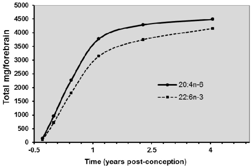
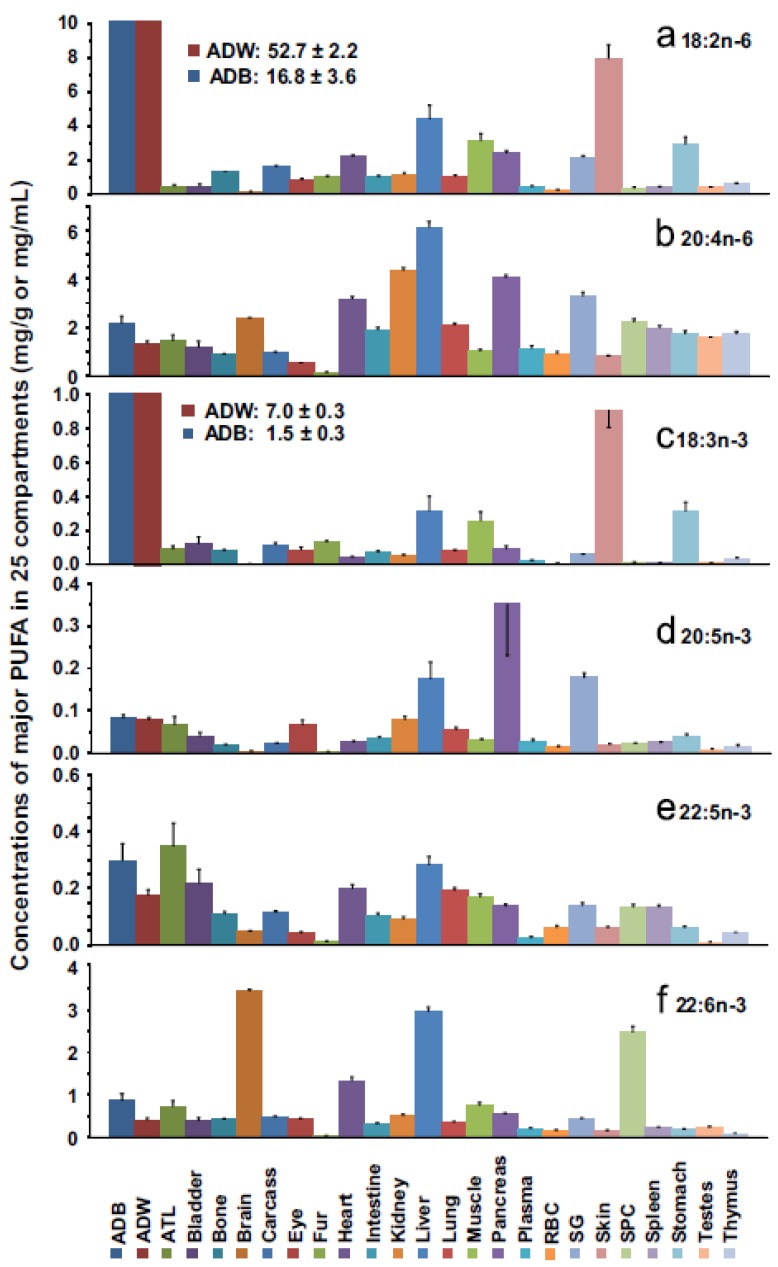
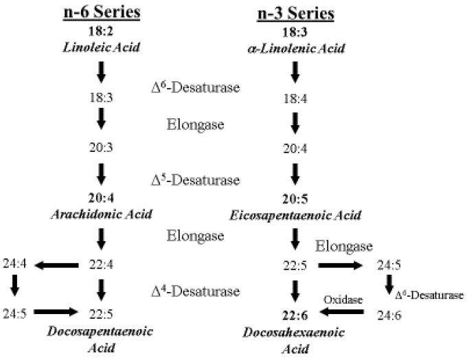
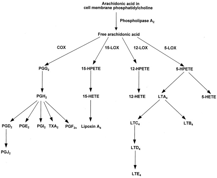
Arachidonic acid (ARA, 20:4n-6) is an n-6 polyunsaturated 20-carbon fatty acid formed by the biosynthesis from linoleic acid (LA, 18:2n-6). This review considers the essential role that ARA plays in infant development. ARA is always present in human milk at a relatively fixed level and is accumulated in tissues throughout the body where it serves several important functions. Without the provision of preformed ARA in human milk or infant formula the growing infant cannot maintain ARA levels from synthetic pathways alone that are sufficient to meet metabolic demand. During late infancy and early childhood the amount of dietary ARA provided by solid foods is low. ARA serves as a precursor to leukotrienes, prostaglandins, and thromboxanes, collectively known as eicosanoids which are important for immunity and immune response. There is strong evidence based on animal and human studies that ARA is critical for infant growth, brain development, and health. These studies also demonstrate the importance of balancing the amounts of ARA and DHA as too much DHA may suppress the benefits provided by ARA. Both ARA and DHA have been added to infant formulas and follow-on formulas for more than two decades. The amounts and ratios of ARA and DHA needed in infant formula are discussed based on an in depth review of the available scientific evidence.

Keywords: arachidonic acid; docosahexaenoic acid; growth; human milk; infant formula; long-chain polyunsaturated fatty acids.


Figures

Figure 1
Figure 1
Long-chain polyunsaturated fatty acids (LCPUFA) accretion in the human brain during perinatal development (Data from Martinez [15]).
Figure 2
Figure 2
Distribution of fatty acids in 25 different tissue compartments in young male rats. Abbreviations: ATL, adrenal gland, thyroid gland, mandibular gland, and lymph nodes; RBC, red blood cell; SG, salivary gland; ADB, brown adipose tissue; ADW, white adipose tissue (from Salem et al. [52]).
Figure 3
Figure 3
Metabolic pathways of linoleic and α-linolenic acid (Adapted from Lauritzen et al. [10]).
Figure 4
Figure 4
Generalized pathway for the conversion of ARA to eicosanoids. COX, cyclooxygenase; HETE, hydroxyeicosatetraenoic acid; HPETE, hydroperoxyeicosatetraenoic acid; LOX, lipoxygenase; LT, leukotriene; PG, prostaglandin; TX, thromboxane (from Calder [98]).
Figure 5
Figure 5
The role of ARA in bone development and homeostatic regulation of vitamin D3 and parathyroid hormone (PTH) levels along the parathyroid gland-kidney axis during growth. ARA and vitamin D3 are acquired from the diet and/or from endogenous sources. ARA mediates vitamin D3 regulation of chondrocyte proliferation and growth plate mineralization during bone elongation. As vitamin D3 is metabolized and levels subside, ARA-dependent PTH suppression is diminished and PTH production by the parathyroid gland is upregulated. This results in increased periosteal bone mineral content (appositional bone growth). In kidney, PTH induces the ARA-mediated increase in vitamin D3 activation and secretion, elevating the amount of vitamin D3 in circulation. The cycle continues as the restoration of vitamin D3 results in the ARA-dependent suppression of PTH and stimulates longitudinal bone growth.
Figure 6
Figure 6
Kaplan-Meier survival curves of Fads1 mice, AA = ARA. (A) Fads1 null mice exhibited low viability when fed a standard AA-free diet; n = 37 for wild-type, n = 44 for heterozygous, n = 11 for Null; (B) Dietary supplementation with AA (0.1% and 0.4%, w/w) partially reversed the Fads1 null mouse phenotype; n = 5 for Null + 0.1% AA, n = 3 for Null + 0.4% AA. Supplementation with 2.0% AA completely reverse the Null phenotype; n = 4 for Null ± 2% AA (from Fan et al. [191]).
Figure 7
Figure 7
Schematic summary of molecular events and functional outcomes involved in metabolism of ARA. ARA is derived from endogenous synthesis or directly from the diet and is incorporated into cellular membrane complex lipids. Within the lipid bilayer, ARA is enriched in PE and PI in the inner membrane. Coordination of spatial-temporal interactions between molecular and cellular components and activities are mediated by metabolites of, or molecules associated with metabolism of ARA. Metabolism of ARA is triggered by activation of transmembrane receptors as a result of binding a ligand. A few examples of receptor-mediated activation of ARA metabolism include glucose, vitamin D3, Ca2+, or antigen presentation or detection by immune cells. ARA released from the membrane by the actions of PLA2 or metabolized by enzymes such as COX, CYP450, and/or LOX can act directly or serve as a substrate for various enzymes to produce second-messengers. ARA-derived eicosanoids, including prostaglandins, leukotrienes, lipoxins, and HETEs regulate numerous activities including passage of ions between subcellular compartments, interactions between various structures or cells, and nuclear regulation of gene transcription by PPARs activators. Within the inner leaflet of cell membranes, ARA is enriched in micro-domains and is involved in regulation of receptor mediated activities. In addition, micro-domains serve as foundations for biophysical interactions between subcellular structures such as microtubules and other cytoskeletal activities including vesicular transport. The consequences of temporal-spatial regulation include coordinated release of hormones, expression of various cell functions, and/or alterations in phenotypes, and cellular motility. Examples of PPAR-regulated gene products involved tissue uptake of LCPUFA and oxidation of stored lipids: MFSD2A, major facilitator of superfamily domain-containing protein 2A. Membrane components: Chol, cholesterol; Gang, gangliosides; PC, phosphatidylcholine; PE, phosphatidylethanolamine; PI, phosphatidylinositol; PS, phosphatidylserine; SPM, Sphingomyelin. Nuclear transcription factors: PPAR, peroxisome-proliferator activator receptors; RXR, retinoid X receptors. TM, transmembrane receptors. Enzymes: COX, cyclooxygenase; CYP450, cytochrome P450; LOX, lipoxygenase; PLA2, Phospholipase A2; PLC, Phospholipase C. Signaling molecules: PG, prostaglandin (Adapted from Pike [32]).

References

    1. Martinez M. Tissue levels of polyunsaturated fatty acids during early human development. J. Pediatr. 1992;120:S129–S138. doi: 10.1016/S0022-3476(05)81247-8. - DOI - PubMed
    1. Koletzko B., Carlson S.E., van Goudoever J.B. Should infant formula provide both omega-3 DHA and omega-6 arachidonic acid? Ann. Nutr. Metab. 2015;66:137–138. doi: 10.1159/000377643. - DOI - PubMed
    1. Bolling K. Infant Feeding Survey, 2005. [(accessed on 17 August 2015)]; Available online: http://www.hscic.gov.uk/pubs/ifs2005.
    1. Centers for Disease Control and Prevention, Division of Nutrition, Physical Activity, and Obesity Breastfeeding Report Card. [(accessed on 18 August 2015)];2014 Available online: http://www.cdc.gov/breastfeeding/data/reportcard.htm.
    1. Brenna J.T., Varamini B., Jensen R.G., Diersen-Schade D.A., Boettcher J.A., Arterburn L.M. Docosahexaenoic and arachidonic acid concentrations in human milk worldwide. Am. J. Clin. Nutr. 2007;85:1457–1464. - PubMed

Publication types