Review
. 2016 Apr 11;5(4):45.
doi: 10.3390/jcm5040045.

Endothelial to Mesenchymal Transition (EndoMT) in the Pathogenesis of Human Fibrotic Diseases

Affiliations
Review

Endothelial to Mesenchymal Transition (EndoMT) in the Pathogenesis of Human Fibrotic Diseases

Sonsoles Piera-Velazquez et al. J Clin Med. .

Abstract

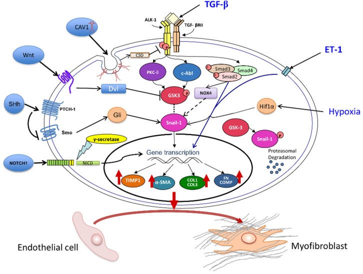
Fibrotic diseases encompass a wide spectrum of clinical entities including systemic fibrotic diseases such as systemic sclerosis, sclerodermatous graft versus host disease, nephrogenic systemic fibrosis, and IgG₄-associated sclerosing disease, as well as numerous organ-specific disorders including radiation-induced fibrosis, and cardiac, pulmonary, liver, and kidney fibrosis. Although their causative mechanisms are quite diverse, these diseases share the common feature of an uncontrolled and progressive accumulation of fibrous tissue macromolecules in affected organs leading to their dysfunction and ultimate failure. The pathogenesis of fibrotic diseases is complex and despite extensive investigation has remained elusive. Numerous studies have identified myofibroblasts as the cells responsible for the establishment and progression of the fibrotic process. Tissue myofibroblasts in fibrotic diseases originate from several sources including quiescent tissue fibroblasts, circulating CD34+ fibrocytes, and the phenotypic conversion of various cell types including epithelial and endothelial cells into activated myofibroblasts. However, the role of the phenotypic transition of endothelial cells into mesenchymal cells (Endothelial to Mesenchymal Transition or EndoMT) in the pathogenesis of fibrotic disorders has not been fully elucidated. Here, we review the evidence supporting EndoMT's contribution to human fibrotic disease pathogenesis.

Keywords: EndoMT; Endothelial Mesenchymal Transition; collagen; endothelial cell; extracellular matrix; fibrosis; fibrotic diseases; idiopathic pulmonary fibrosis; myofibroblast; systemic sclerosis; transforming growth factor-β.


Figures

Figure 1
Figure 1
Molecular Mechanisms of EndoMT. The diagram shows the TGF-β, ET-1, NOTCH, CAV-1, Wnt, NOX4, and HIF-1α pathways that may participate in the EndoMT process. The most important pathway is initiated following TGF-β-binding and subsequent activation of Smad-dependent and Smad-independent TGF-β intracellular signaling. TGF-β causes a direct stimulation of NOX4 expression that results in Snail1 mediated EndoMT. ET-1 induces a synergistic stimulation of TGF-β-induced EndoMT involving the canonical Smad pathways. Hypoxia also induces EndoMT through the effects of Hif1α activation of Snail1. Snail1 has emerged as a crucial regulatory molecule in EndoMT and its levels are modulated by GSK3-mediated phosphorylation as phosphorylated Snail1 undergoes proteasomal degradation. Cav1 exerts an inhibitory effect owing to the internalization of TGF-β receptors and their subsequent degradation. Morphogen pathways including Wnt, Sonic Hh, and NOTCH also may modulate EndoMT. The ultimate effect of these complex intracellular signaling events is the activation of a mesenchymal cell specific transcriptional gene regulation program leading to the increased production of various myofibroblast-specific and profibrotic macromolecules including α-SMA, COL1, COL3, FN, COMP, and the MMP-inhibitor TIMP. These events are accompanied by the repression of EC-specific gene products such as CD31/PECAM-1, VE-cadherin, and von Willebrand Factor (not shown in the diagram) resulting in the phenotypic conversion of EC into myofibroblasts, the cells ultimately responsible for the fibrotic process.
Figure 2
Figure 2
Immunohistology and confocal microscopy staining of medium-sized pulmonary arteries in SSc-associated pulmonary fibrosis lung tissues. (A) Histopathology of a pulmonary arteriole showing severe proliferative vasculopathy with luminal occlusion; (B) CD31-expressing cells (brown staining) in the subendothelial region of a small pulmonary arteriole and in the lung parenchyma (red circles). (CE) small arteriole in affected SSc lung; (C) Staining for vWF (green); (D) Staining for α-SMA (red); (E) Overlay (yellow). Note numerous cells in the endothelial lining (arrows) and one cell in the subendothelial tissue (arrowhead) displaying co-expression of EC (vWF) and myofibroblast (α-SMA) molecular markers as evidenced by the yellow color in the overlay image. Adapted from Ref. [64].

References

    1. Rockey D.C., Bell P.D., Hill J.A. Fibrosis—A common pathway to organ injury and failure. N. Engl. J. Med. 2015;372:1138–1149. - PubMed
    1. Rosenbloom J., Castro S.V., Jimenez S.A. Fibrotic Diseases: Cellular and Molecular Mechanisms and Novel Therapies. Ann. Int. Med. 2010;152:159–166. - PubMed
    1. Wynn T.A. Common and unique mechanisms regulate fibrosis in various fibroproliferative diseases. J. Clin. Invest. 2007;117:524–529. doi: 10.1172/JCI31487. - DOI - PMC - PubMed
    1. Varga J., Abraham D. Systemic sclerosis: A prototypic multisystem fibrotic disorder. J. Clin. Invest. 2007;117:557–567. doi: 10.1172/JCI31139. - DOI - PMC - PubMed
    1. Gabrielli A., Avvedimento E.V., Krieg T. Scleroderma. N. Engl. J. Med. 2009;360:1989–2003. doi: 10.1056/NEJMra0806188. - DOI - PubMed

LinkOut - more resources