. 2016 Apr 13;19(4):515-528.
doi: 10.1016/j.chom.2016.03.001.

NLRX1 Sequesters STING to Negatively Regulate the Interferon Response, Thereby Facilitating the Replication of HIV-1 and DNA Viruses

Affiliations

NLRX1 Sequesters STING to Negatively Regulate the Interferon Response, Thereby Facilitating the Replication of HIV-1 and DNA Viruses

Haitao Guo et al. Cell Host Microbe. .

Abstract

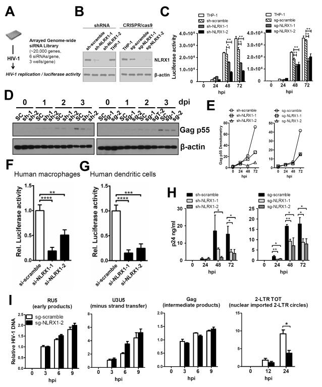
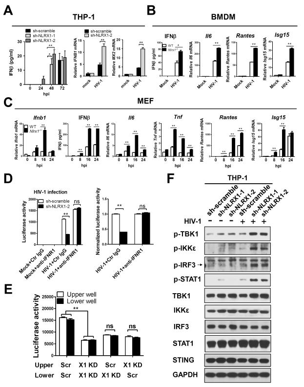
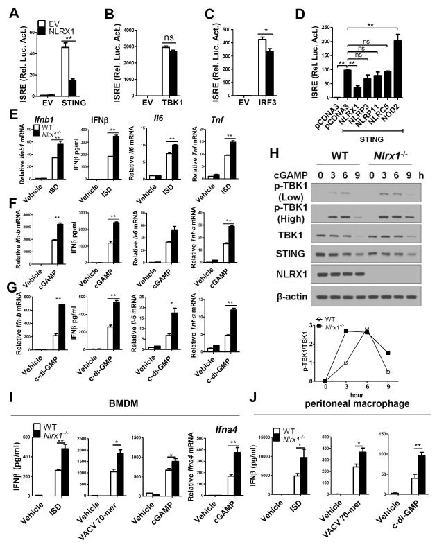
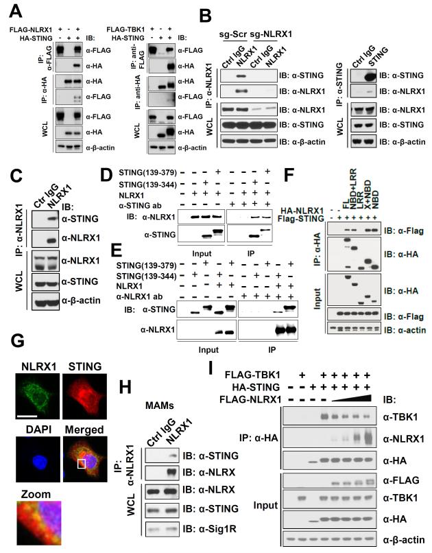
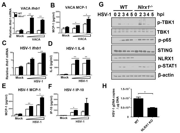
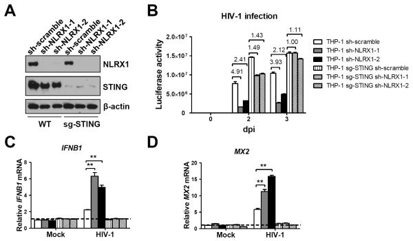
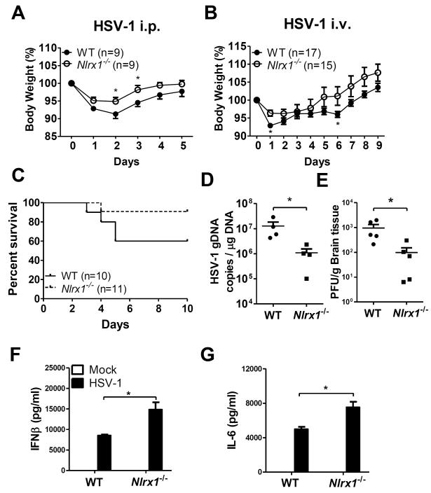
Understanding the negative regulators of antiviral immune responses will be critical for advancing immune-modulated antiviral strategies. NLRX1, an NLR protein that negatively regulates innate immunity, was previously identified in an unbiased siRNA screen as required for HIV infection. We find that NLRX1 depletion results in impaired nuclear import of HIV-1 DNA in human monocytic cells. Additionally, NLRX1 was observed to reduce type-I interferon (IFN-I) and cytokines in response to HIV-1 reverse-transcribed DNA. NLRX1 sequesters the DNA-sensing adaptor STING from interaction with TANK-binding kinase 1 (TBK1), which is a requisite for IFN-1 induction in response to DNA. NLRX1-deficient cells generate an amplified STING-dependent host response to cytosolic DNA, c-di-GMP, cGAMP, HIV-1, and DNA viruses. Accordingly, Nlrx1(-/-) mice infected with DNA viruses exhibit enhanced innate immunity and reduced viral load. Thus, NLRX1 is a negative regulator of the host innate immune response to HIV-1 and DNA viruses.


Figures

Figure 1
Figure 1. NLRX1 is required for HIV-1 infection
(A) Scheme of unbiased screening of host factors required for HIV-1 infection which identified that siRNA targeting NLRX1 reduced HIV-1 infection. (B) Knockdown of NLRX1 in THP-1 cells by shRNA (left) or CRISPR-Cas9/sgRNA (right). (C-E) THP-1 cells with scrambled or shRNA or sgRNA targeting NLRX1 were infected with a single-cycle luciferase reporter virus pseudo-typed with a pH-dependent envelope VSV-G (HIV-VSV). After 24, 48, and 72 hours, luciferase activity (C) and the expression of HIV-1 Gag p55 protein (D) were determined. Densitometry analysis was performed for Gag p55 levels shown in panel D (E). SC: scrambled control, sh-1: sh-NLRX1-1, sh-2, sh-NLRX1-2, sg-1: sg-NLRX1-1, sg-2: sg-NLRX1-2. (F and G) Knockdown of NLRX1 decreases HIV-1 infection in human primary macrophages (F) and dendritic cells (G). Monocyte derived macrophage and monocyte derived dendritic cells were transfected with scrambled siRNA or siRNA targeting NLRX1 followed by infection with HIV-VSV. HIV-1 infection as reflected by luciferase activity is shown. Data pooled from three or four different donors. (H) Same as C except that the infection was done by using a CCR5/CXCR4 dual tropic HIV-1 strain R3A. HIV-1 particle release was quantified by p24 ELISA. (I) Real-Time PCR analysis of HIV-1 reverse transcription and cDNA nuclear import as previously described (Mbisa et al., 2009). THP-1 cells with scrambled or shRNA or sgRNA targeting NLRX1 were infected with a HIV-VSV. At indicated time points, total DNA was extracted and HIV-1 DNA was quantified by amplifying indicated target sequences. Data are presented as the mean ± standard error of the mean (SEM). *p < 0.05, **p < 0.01, ***p < 0.001, ****p < 0.0001 (Student’s t test). All data are representative of two or three independent experiments. See also Figure S1.
Figure 2
Figure 2. Removal of NLRX1 enhances innate immune response upon HIV-1 infection
(A) THP-1 cells with shNLRX1 or scrambled control shRNA were infected with HIV-VSV (MOI=1) and the induction of IFNβ was determined over time. IFNB1 and MX2 mRNA levels were determined by qPCR 18 hpi. (B) WT or Nlrx1−/− BMDM were infected with HIV-VSV (MOI=1), at different time points, IFNβ was measured by ELISA and Il6, Isg15 and Rantes were measured by qPCR. (C) WT or Nlrx1−/− MEF were infected with HIV-VSV, MOI=1, at different time points. Ifnb was measured by qPCR and ELISA and Il6, Tnf, Isg15 and Rantes were measured by qPCR. (D) THP-1 cells with shNLRX1 or scrambled control shRNA were infected with HIV-VSV (MOI=1) in the presence of anti-IFNR1 or control IgG. At 72 hpi, luciferase activity (normalized by total protein) was determined to measure virus infection (left panel). Luciferase data were normalized by setting scrambled control as 1 (right panel). (E) THP-1 cells with shNLRX1 (X1 KD) or scrambled control (Scr) shRNA were infected with HIV-VSV, MOI=1, and seeded in the plate with transwell as indicated, and luciferase activity from upper and lower wells was determined for virus infection 72 hpi. Luciferase activity was normalized by total protein. (F) THP-1 cells with shNLRX1 or scrambled control shRNA were infected with HIV-VSV, MOI=1. 1 day after infection, cells were collected and p-TBK1, p-IKKε, p-IRF3, p-STAT1, TBK-1, IKKε, IRF3, STAT1, and STING were determined by western blot. GAPDH was used as the loading control. Data are presented as the mean ± standard error of the mean (SEM). *p < 0.05, **p < 0.01 (Student’s t test). All data are representative of at least three independent experiments. See also Figure S2.
Figure 3
Figure 3. NLRX1 inhibits STING-dependent DNA sensing pathway
(A-C) HEK293T cells were transfected with the ISRE promoter reporter with the internal control Renilla luciferase reporter pLR-TK. Either empty vector or NLRX1 expression plasmid was transfected. STING (A), TBK1 (B) or IRF3 (C) was used as activator of ISRE reporter as indicated. Luciferase assays were performed 24 hr after transfection. (D) Same as (A), except cells were transfected with either an empty plasmid or with NLRX1, NLRP3, NLRP11, NLRC5 or NOD2 expression plasmid. (E-G) WT and Nlrx1−/− MEFs were transfected with ISD (E), cGAMP (F), or c-di-GMP (G) respectively. Ifnb, Il-6, and Tnf-α transcripts and IFNβ were measured 6 hr after transfection as indicated. (H) Immunoblot of phosphorylated (p-) TBK1, TBK1, STING, and NLRX1 in lysates of WT and Nlrx1−/− MEFs transfected with cGAMP for indicated time points. β-actin was used as the loading control. Densitometry analysis was performed for (p-) TBK1 levels by normalizing to TBK1. (I) WT and Nlrx1−/− BMDMs were transfected by ISD, VACV 70-mer, or cGAMP. IFNβ and Ifna4 transcript were measured 3 hr after transfection. (J) Peritoneal macrophages isolated from WT and NLRX1−/− mice were transfected with ISD, VACV 70-mer, or c-di-GMP, respectively. IFNβ was assessed 4 hr after transfection. Data are presented as the mean ± standard error of the mean (SEM). *p < 0.05, **p < 0.01 (Student’s t test). All data are representative of at least three independent experiments.
Figure 4
Figure 4. NLRX1 disrupts STING-TBK1 interaction
(A) Association of over-expressed NLRX1 and STING (left) and of overexpressed STING and TBK1 (right) in HEK293T cells. FLAG-NLRX1 and HA-STING was singly expressed or co-expressed in HEK293T cells. Co-immunoprecipitaion was performed with anti-FLAG beads or anti-HA beads respectively. Similarly, FLAG-TBK1 and HA-STING was singly expressed or co-expressed in HEK293T cells. Co-immunoprecipitaion was performed with anti-FLAG beads or anti-HA beads respectively. (B) Determination of endogenous NLRX1-STING association in THP-1 cells with scrambled sgRNA (sg-Scr) or sgRNA for NLRX1 (sg-NLRX1). Immunoprecipitation was performed by using anti-NLRX1, anti-STING or control IgG. (C) Determination of endogenous NLRX1-STING association in primary MEF. Immunoprecipitation was performed by using anti-NLRX1 or control IgG. (D and E) Recombinant NLRX1 and STING in vitro binding assay was performed by using antibodies against STING (D) and NLRX1 (E). Immunoblots were performed with the antibodies indicated. (F) Mapping of NLRX1 domains involved in interaction with STING. FLAG tagged full length NLRX1, NLRX1 lacking X domain (NBD+LRR), NLRX1 LRR, NLRX1 lacking LRR domain (X+NBD), and NLRX1 NBD was expressed in the absence or presence of HA-STING in HEK293T cells. Immunoprecipitation was performed by anti-HA beads. (G) Partial co-localization of NLRX1 and STING. FLAG-NLRX1 and HA-STING was co-expressed in HEK293T cells and immunofluorescence staining was performed. Pictures were taken using confocal microscopy. Scale bar equals to 20 μm. (H) Determination of endogenous NLRX1-STING association in MAMs isolated from human foreskin fibroblast (HFF) cells. Immunoprecipitation was performed by using anti-NLRX1 or control IgG. Sig1R was used as the MAMs marker. (I) NLRX1 attenuates STING-TBK1 interaction. STING and TBK1 were co-expressed in HEK293T cells with increasing dose of NLRX1. Immunoprecipitation was performed with anti-HA beads. All data are representative of at least three independent experiments. See also Figure S3-S7.
Figure 5
Figure 5. Nlrx1 deficiency causes enhanced innate immune response to DNA virus infection
(A and B) WT and Nlrx1−/− MEFs were infected with vaccinia virus (VACA) (MOI=1 or 5), Ifnb transcript (E) and MCP-1 (F) were assessed. (C-F) WT and Nlrx1−/− MEFs were infected with HSV-1 (MOI=0.1 or 1), Ifnb transcript (C), IL-6 (D), MCP-1 (E), and IP-10 (F) were assessed. (G) Immunoblot of phosphorylated (p-) TBK1, TBK1, (p-) STAT1, (p-) p65, STING, and NLRX1 in lysates of WT and Nlrx1−/− BMDMs infected with HSV-1 (MOI=1) for indicated time points. β-actin was used as the loading control. (H) WT or Nlrx1−/− BMDMs were infected with HSV-1 (MOI=0.1) and HSV-1 genome copies were quantified by qPCR 24 hr post infection. Data are presented as the mean ± standard error of the mean (SEM). *p < 0.05, **p < 0.01 (Student’s t test). Data in A-F are from one experiment and data in G and H are representative of at least three independent experiments.
Figure 6
Figure 6. NLRX1 promotes HIV-1 infection in a STING-dependent manner
(A) NLRX1 and STING expression were attenuated by transducing THP-1 cells with lentivirus expressing shRNA for NLRX1 and Cas9 nuclease plus sgRNA targeting STING. Attenuation of NLRX1 and STING expression were confirmed by immunoblot. (B) THP-1 cells containing scrambled shRNA, shRNA for NLRX1, or both shRNA for NLRX1 and sgRNA for STING were infected with HIV-VSV, MOI=1, luciferase activity was determined 2 and 3 dpi. Fold differences between indicated groups were labeled on top of each group. (C and D). Same as (B) except that IFNB1 (C) and MX2 (D) mRNA levels were determined by qPCR 16 hpi. Data for luciferase and qPCR are presented as the mean ± standard error of the mean (SEM). *p < 0.05, **p < 0.01 (Student’s t test). Data are representative of at least three independent experiments.
Figure 7
Figure 7. Nlrx1 deficient mice are more resistant to HSV-1 infection
(A) WT and Nlrx1−/− mice were infected i.p. with HSV-1 (2×10⁷ pfu) and body weight was monitored. (B-G) WT and Nlrx1−/− mice were infected i.v. with HSV-1. Body weight (B), percentage of survival (C), HSV-1 genomic DNA copies (D), viral titers in the brain (E), IFNβ (F) and IL-6 (G) in the serum were assessed. In B, D, E, F and G, the dose of infection is 2×10⁷ pfu/mouse and in C, the dose of infection is 5×10⁷ pfu/mouse. *p < 0.05 (Student’s t test or Mann-Whitney U test). Data are representative of at least two independent experiments.

References

    1. Abdul-Sater AA, Said-Sadier N, Lam VM, Singh B, Pettengill MA, Soares F, Tattoli I, Lipinski S, Girardin SE, Rosenstiel P, et al. Enhancement of reactive oxygen species production and chlamydial infection by the mitochondrial Nod-like family member NLRX1. The Journal of biological chemistry. 2010;285:41637–41645. - PMC - PubMed
    1. Allen IC. Non-Inflammasome Forming NLRs in Inflammation and Tumorigenesis. Frontiers in immunology. 2014;5:169. - PMC - PubMed
    1. Allen IC, Moore CB, Schneider M, Lei Y, Davis BK, Scull MA, Gris D, Roney KE, Zimmermann AG, Bowzard JB, et al. NLRX1 protein attenuates inflammatory responses to infection by interfering with the RIG-I-MAVS and TRAF6-NF-kappaB signaling pathways. Immunity. 2011;34:854–865. - PMC - PubMed
    1. Altfeld M, Gale M., Jr. Innate immunity against HIV-1 infection. Nature immunology. 2015;16:554–562. - PubMed
    1. Burdette DL, Monroe KM, Sotelo-Troha K, Iwig JS, Eckert B, Hyodo M, Hayakawa Y, Vance RE. STING is a direct innate immune sensor of cyclic di-GMP. Nature. 2011;478:515–518. - PMC - PubMed

Publication types

MeSH terms