. 2016 Aug;23(8-9):649-656.
doi: 10.1038/gt.2016.35. Epub 2016 May 12.

Vascular endothelial growth factor-A gene electrotransfer promotes angiogenesis in a porcine model of cardiac ischemia

Affiliations

Vascular endothelial growth factor-A gene electrotransfer promotes angiogenesis in a porcine model of cardiac ischemia

Anna A Bulysheva et al. Gene Ther. 2016 Aug.

Abstract

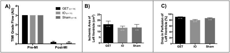
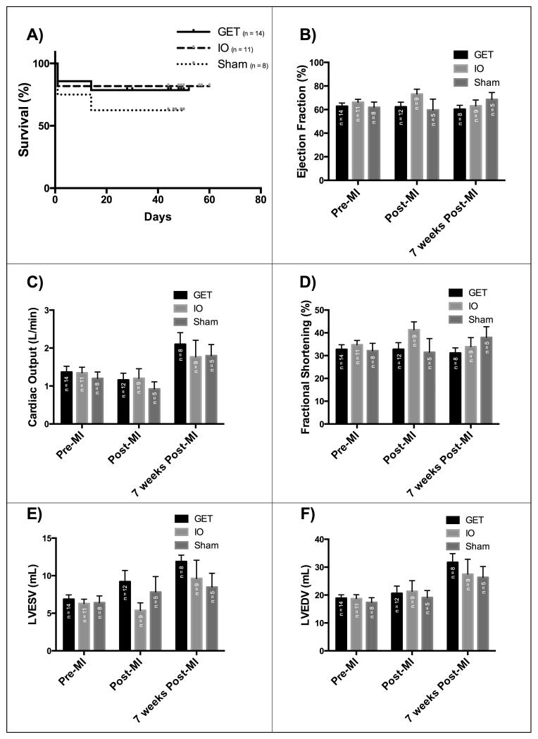
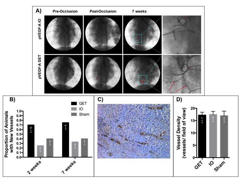
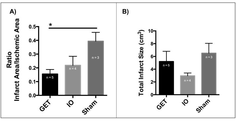
This study aimed to assess safety and therapeutic potential of gene electrotransfer (GET) as a method for delivery of plasmid encoding vascular endothelial growth factor A (VEGF-A) to ischemic myocardium in a porcine model. Myocardial ischemia was induced by surgically occluding the left anterior descending coronary artery in swine. GET following plasmid encoding VEGF-A injection was performed at four sites in the ischemic region. Control groups either received injections of the plasmid without electrotransfer or injections of the saline vehicle. Animals were monitored for 7 weeks and the hearts were evaluated for angiogenesis, myocardial infarct size and left ventricular contractility. Arteriograms suggest growth of new arteries as early as 2 weeks after treatment in electrotransfer animals. There is a significant reduction of infarct area and left ventricular contractility is improved in GET-treated group compared with controls. There was no significant difference in mortality of animals treated with GET of plasmid encoding VEGF-A from the control groups. Gene delivery of plasmid encoding VEGF-A to ischemic myocardium in a porcine model can be accomplished safely with potential for myocardial repair and regeneration.


Figures

Figure 1
Figure 1. Permanent ischemia was induced via LAD occlusion
A) TIMI flow index drops significantly after occlusion, with no significant difference in TIMI between treatment groups (Two-way ANOVA and Tukey’s multiple comparisons test, alpha=0.05, P = <0.0001). B) SPY based measurement of ischemic surface area of the left ventricle indicates consistent occlusion between treatment groups, with no significant difference in ischemic area (One-way ANOVA, alpha = 0.05, P=0.6388). C) SPY based measurement of drop in perfusion in the ischemic area of the left ventricle indicates a consistent reduction in perfusion, with GET group receiving a slightly larger drop in perfusion than the IO group (One-way ANOVA, Tukey’s multiple comparisons test alpha=0.05, P=0.0012).
Figure 2
Figure 2. GET of VEGF-A has no deleterious effects on survival or heart performance over 7 weeks post MI and gene delivery
A) Kaplan-Meier Survival curve with no significant difference between the groups (Logrank test for trend, P=0.4639), B) Ejection fraction (P=0.9240), C) Cardiac Output (P=0.5603), D) Fractional Shortening(P=0.3775), E) Left ventricular end systolic volume (P=0.5603), F) Left Ventricular end diastolic volume(P=0.92), with no significant difference between treatment groups based on echocardiographic analysis (Two-way ANOVA and Tukey’s multiple comparison test, alpha=0.05)
Figure 3
Figure 3. GET of pVEGF-A induces angiogenesis in ischemic myocardium at treatment sites
A) Arteriograms of GET and IO animals, before occlusion, immediately after occlusion and 7 weeks after occlusion, with new arteries visible in the GET heart. *LAD occlusion site, red arrows point to new vessels. B) The proportion of hearts with new arteries visible via arteriogram 2 (P=0.152313) and 7 weeks (P=0.243917) after treatment is higher in hearts treated with GET of pVEGF-A (Chi-square test). C) vWF staining for blood vessels at a treatment site of GET of pVEGF-A seven weeks after MI and treatment. D) Blood vessel density at treatment sites seven weeks after MI and treatment is not significantly different between treatment groups on a microscopic level (One-way ANOVA, alpha=0.05, P=0.9704).
Figure 4
Figure 4. Myocardial infarct size is reduced at GET of pVEGF treatment sites 7 weeks post treatment
A) Infarct area to ischemic area ratio is significantly lower in GET treated left ventricles than in Sham control group, (One-way ANOVA, and Tukey’s multiple comparison test, alpha=0.05, p=0.0349), B) Total myocardial infarct size including untreated septum and untreated right ventricle, with no significant differences between the groups (One-way ANOVA, alpha=0.05, P=0.2028).

References

    1. Go AS, Mozaffarian D, Roger VL, Benjamin EJ, Berry JD, Borden WB, et al. Heart disease and stroke statistics--2013 update: a report from the American Heart Association. Circulation. 2013;127:e6–e245. - PMC - PubMed
    1. 2012 NHLBI Morbidity and Mortality Chart Book. 2013;2014
    1. Krumholz HM, Merrill AR, Schone EM, Schreiner GC, Chen J, Bradley EH, et al. Patterns of hospital performance in acute myocardial infarction and heart failure 30-day mortality and readmission. Circ Cardiovasc Qual Outcomes. 2009;2:407–413. - PubMed
    1. Ross R. The pathogenesis of atherosclerosis: a perspective for the 1990s. Nature. 1993;362:801–809. - PubMed
    1. Mukherjee D, Bhatt DL, Roe MT, Patel V, Ellis SG. Direct myocardial revascularization and angiogenesis--how many patients might be eligible? Am J Cardiol. 1999;84:598–600. A8. - PubMed

Publication types

MeSH terms

Substances