

The association of motor imagery and kinesthetic illusion prolongs the effect of transcranial direct current stimulation on corticospinal tract excitability
- PMID: 27079199
- PMCID: PMC4832525
- DOI: 10.1186/s12984-016-0143-8
The association of motor imagery and kinesthetic illusion prolongs the effect of transcranial direct current stimulation on corticospinal tract excitability
Abstract
Background: A kinesthetic illusion induced by a visual stimulus (KI) can produce vivid kinesthetic perception. During KI, corticospinal tract excitability increases and results in the activation of cerebral networks. Transcranial direct current stimulation (tDCS) is emerging as an alternative potential therapeutic modality for a variety of neurological and psychiatric conditions, such that identifying factors that enhance the magnitude and duration of tDCS effects is currently a topic of great scientific interest. This study aimed to establish whether the combination of tDCS with KI and sensory-motor imagery (MI) induces larger and longer-lasting effects on the excitability of corticomotor pathways in healthy Japanese subjects.
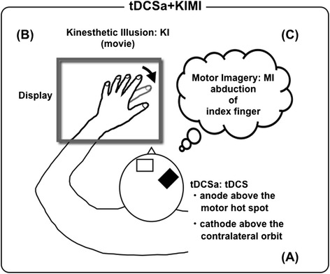
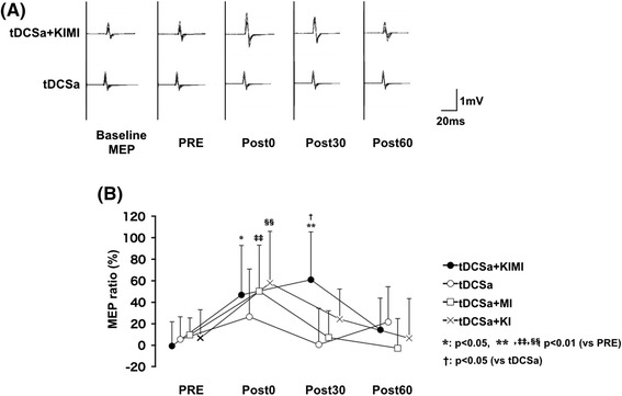
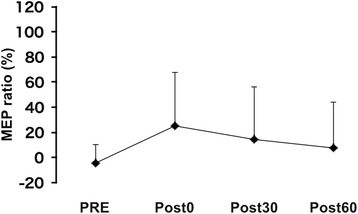
Methods: A total of 21 healthy male volunteers participated in this study. Four interventions were investigated in the first experiment: (1) anodal tDCS alone (tDCSa), (2) anodal tDCS with visually evoked kinesthetic illusion (tDCSa + KI), (3) anodal tDCS with motor imagery (tDCSa + MI), and (4) anodal tDCS with kinesthetic illusion and motor imagery (tDCSa + KIMI). In the second experiment, we added a sham tDCS intervention with kinesthetic illusion and motor imagery (sham + KIMI) as a control for the tDCSa + KIMI condition. Direct currents were applied to the right primary motor cortex. Corticospinal excitability was examined using transcranial magnetic stimulation of the area associated with the left first dorsal interosseous.
Results: In the first experiment, corticomotor excitability was sustained for at least 30 min following tDCSa + KIMI (p < 0.01). The effect of tDCSa + KIMI on corticomotor excitability was greater and longer-lasting than that achieved in all other conditions. In the second experiment, significant effects were not achieved following sham + KIMI.
Conclusions: Our results suggest that tDCSa + KIMI has a greater therapeutic potential than tDCS alone for inducing higher excitability of the corticospinal tract. The observed effects may be related to sustained potentiation of resultant cerebral activity during combined KI, MI, and tDCSa.
Keywords: Corticospinal tract; Kinesthetic illusion; Motor imagery; Transcranial direct current stimulation; Transcranial magnetic stimulation; Visual stimulation.
Figures
Comment in
-
Enhancing transcranial direct current stimulation via motor imagery and kinesthetic illusion: crossing internal and external tools.J Neuroeng Rehabil. 2016 Jun 1;13(1):50. doi: 10.1186/s12984-016-0156-3. J Neuroeng Rehabil. 2016. PMID: 27246465 Free PMC article.
Similar articles
-
Time course of changes in corticospinal excitability induced by motor imagery during action observation combined with peripheral nerve electrical stimulation.Exp Brain Res. 2019 Mar;237(3):637-645. doi: 10.1007/s00221-018-5454-5. Epub 2018 Dec 8. Exp Brain Res. 2019. PMID: 30536148
-
Transcranial direct-current stimulation reduced the excitability of diaphragmatic corticospinal pathways whatever the polarity used.Respir Physiol Neurobiol. 2013 Oct 1;189(1):183-7. doi: 10.1016/j.resp.2013.07.024. Epub 2013 Aug 7. Respir Physiol Neurobiol. 2013. PMID: 23933029 Clinical Trial.
-
Slow-oscillatory transcranial direct current stimulation can induce bidirectional shifts in motor cortical excitability in awake humans.Neuroscience. 2010 Apr 14;166(4):1219-25. doi: 10.1016/j.neuroscience.2010.01.019. Epub 2010 Jan 18. Neuroscience. 2010. PMID: 20083166
-
Does cerebellar non-invasive brain stimulation affect corticospinal excitability in healthy individuals? A systematic review of literature and meta-analysis.Neurosci Lett. 2019 Jul 27;706:128-139. doi: 10.1016/j.neulet.2019.05.025. Epub 2019 May 15. Neurosci Lett. 2019. PMID: 31102706
-
Sham transcranial electrical stimulation and its effects on corticospinal excitability: a systematic review and meta-analysis.Rev Neurosci. 2018 Feb 23;29(2):223-232. doi: 10.1515/revneuro-2017-0026. Rev Neurosci. 2018. PMID: 28889119
Cited by
-
Feasibility Case Study for Treating a Patient with Sensory Ataxia Following a Stroke with Kinesthetic Illusion Induced by Visual Stimulation.Prog Rehabil Med. 2020 Oct 28;5:20200025. doi: 10.2490/prm.20200025. eCollection 2020. Prog Rehabil Med. 2020. PMID: 33134593 Free PMC article.
-
Model-Based Analyses for the Causal Relationship Between Post-stroke Impairments and Functional Brain Connectivity Regarding the Effects of Kinesthetic Illusion Therapy Combined With Conventional Exercise.Front Syst Neurosci. 2022 Jan 10;15:804263. doi: 10.3389/fnsys.2021.804263. eCollection 2021. Front Syst Neurosci. 2022. PMID: 35173590 Free PMC article.
-
A Case Series Clinical Trial of a Novel Approach Using Augmented Reality That Inspires Self-body Cognition in Patients With Stroke: Effects on Motor Function and Resting-State Brain Functional Connectivity.Front Syst Neurosci. 2019 Dec 17;13:76. doi: 10.3389/fnsys.2019.00076. eCollection 2019. Front Syst Neurosci. 2019. PMID: 31920571 Free PMC article.
-
Promising Effect of Visually-Assisted Motor Imagery Against Arthrogenic Muscle Inhibition - A Human Experimental Pain Study.J Pain Res. 2021 Feb 3;14:285-295. doi: 10.2147/JPR.S282736. eCollection 2021. J Pain Res. 2021. PMID: 33568937 Free PMC article.
-
Enhancing transcranial direct current stimulation via motor imagery and kinesthetic illusion: crossing internal and external tools.J Neuroeng Rehabil. 2016 Jun 1;13(1):50. doi: 10.1186/s12984-016-0156-3. J Neuroeng Rehabil. 2016. PMID: 27246465 Free PMC article.
References
Publication types
MeSH terms
LinkOut - more resources
Full Text Sources
Other Literature Sources
Medical
