

Autophagy dysfunction and regulatory cystatin C in macrophage death of atherosclerosis
- PMID: 27079462
- PMCID: PMC4988293
- DOI: 10.1111/jcmm.12859
Autophagy dysfunction and regulatory cystatin C in macrophage death of atherosclerosis
Abstract
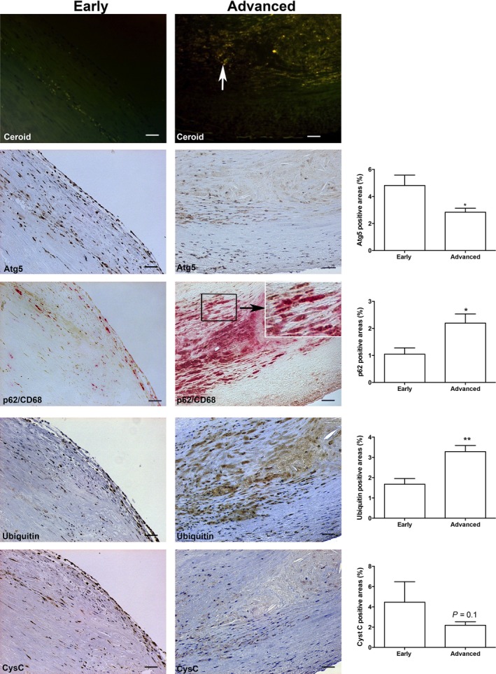
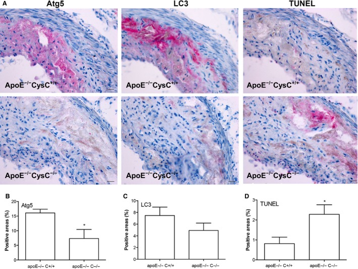
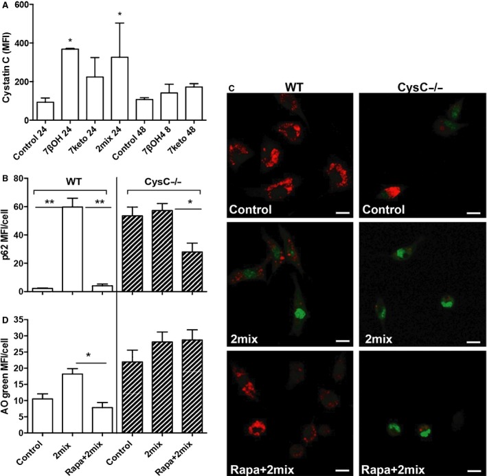
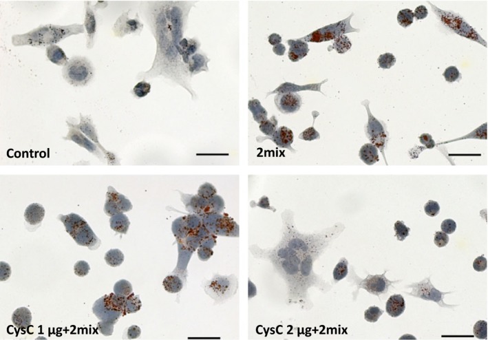
Autophagy dysfunction in mouse atherosclerosis models has been associated with increased lipid accumulation, apoptosis and inflammation. Expression of cystatin C (CysC) is decreased in human atheroma, and CysC deficiency enhances atherosclerosis in mice. Here, we first investigated the association of autophagy and CysC expression levels with atheroma plaque severity in human atherosclerotic lesions. We found that autophagy proteins Atg5 and LC3β in advanced human carotid atherosclerotic lesions are decreased, while markers of dysfunctional autophagy p62/SQSTM1 and ubiquitin are increased together with elevated levels of lipid accumulation and apoptosis. The expressions of LC3β and Atg5 were positively associated with CysC expression. Second, we investigated whether CysC expression is involved in autophagy in atherosclerotic apoE-deficient mice, demonstrating that CysC deficiency (CysC(-/-) ) in these mice results in reduction of Atg5 and LC3β levels and induction of apoptosis. Third, macrophages isolated from CysC(-/-) mice displayed increased levels of p62/SQSTM1 and higher sensitivity to 7-oxysterol-mediated lysosomal membrane destabilization and apoptosis. Finally, CysC treatment minimized oxysterol-mediated cellular lipid accumulation. We conclude that autophagy dysfunction is a characteristic of advanced human atherosclerotic lesions and is associated with reduced levels of CysC. The deficiency of CysC causes autophagy dysfunction and apoptosis in macrophages and apoE-deficient mice. The results indicate that CysC plays an important regulatory role in combating cell death via the autophagic pathway in atherosclerosis.
Keywords: autophagy; cystatin C; lysosomal membrane permeabilization; macrophage cell death.
© 2016 The Authors. Journal of Cellular and Molecular Medicine published by John Wiley & Sons Ltd and Foundation for Cellular and Molecular Medicine.
Figures
Similar articles
-
Lack of the cysteine protease inhibitor cystatin C promotes atherosclerosis in apolipoprotein E-deficient mice.Arterioscler Thromb Vasc Biol. 2005 Oct;25(10):2151-6. doi: 10.1161/01.ATV.0000179600.34086.7d. Epub 2005 Jul 28. Arterioscler Thromb Vasc Biol. 2005. PMID: 16051881
-
Mst1 participates in the atherosclerosis progression through macrophage autophagy inhibition and macrophage apoptosis enhancement.J Mol Cell Cardiol. 2016 Sep;98:108-16. doi: 10.1016/j.yjmcc.2016.08.002. Epub 2016 Aug 3. J Mol Cell Cardiol. 2016. PMID: 27496379
-
Autophagy links inflammasomes to atherosclerotic progression.Cell Metab. 2012 Apr 4;15(4):534-44. doi: 10.1016/j.cmet.2012.02.011. Epub 2012 Mar 20. Cell Metab. 2012. PMID: 22440612 Free PMC article.
-
Autophagy and Its Association with Macrophages in Clonal Hematopoiesis Leading to Atherosclerosis.Int J Mol Sci. 2025 Apr 1;26(7):3252. doi: 10.3390/ijms26073252. Int J Mol Sci. 2025. PMID: 40244103 Free PMC article. Review.
-
Macrophage proliferation and apoptosis in atherosclerosis.Curr Opin Lipidol. 2012 Oct;23(5):429-38. doi: 10.1097/MOL.0b013e328357a379. Curr Opin Lipidol. 2012. PMID: 22964992 Review.
Cited by
-
Autophagy and its regulation by ginseng components.J Ginseng Res. 2019 Jul;43(3):349-353. doi: 10.1016/j.jgr.2018.12.011. Epub 2019 Jan 2. J Ginseng Res. 2019. PMID: 31308805 Free PMC article. Review.
-
Cystatin C Deficiency Increases LPS-Induced Sepsis and NLRP3 Inflammasome Activation in Mice.Cells. 2021 Aug 12;10(8):2071. doi: 10.3390/cells10082071. Cells. 2021. PMID: 34440840 Free PMC article.
-
Macrophage Phenotyping in Atherosclerosis by Proteomics.Int J Mol Sci. 2023 Jan 30;24(3):2613. doi: 10.3390/ijms24032613. Int J Mol Sci. 2023. PMID: 36768933 Free PMC article. Review.
-
Programmed cell death in spinal cord injury pathogenesis and therapy.Cell Prolif. 2021 Mar;54(3):e12992. doi: 10.1111/cpr.12992. Epub 2021 Jan 27. Cell Prolif. 2021. PMID: 33506613 Free PMC article. Review.
-
Stefin B Inhibits NLRP3 Inflammasome Activation via AMPK/mTOR Signalling.Cells. 2023 Nov 29;12(23):2731. doi: 10.3390/cells12232731. Cells. 2023. PMID: 38067160 Free PMC article.
References
-
- Mitra AK, Dhume AS, Agrawal DK. “Vulnerable plaques”–ticking of the time bomb. Can J Physiol Pharmacol. 2004; 82: 860–71. - PubMed
-
- Lougheed M, Moore ED, Scriven DR, et al Uptake of oxidized LDL by macrophages differs from that of acetyl LDL and leads to expansion of an acidic endolysosomal compartment. Arterioscler Thromb Vasc Biol. 1999; 19: 1881–90. - PubMed
-
- Cox BE, Griffin EE, Ullery JC, et al Effects of cellular cholesterol loading on macrophage foam cell lysosome acidification. J Lipid Res. 2007; 48: 1012–21. - PubMed
-
- Li W, Yuan XM, Olsson AG, et al Uptake of oxidized LDL by macrophages results in partial lysosomal enzyme inactivation and relocation. Arterioscler Thromb Vasc Biol. 1998; 18: 177–84. - PubMed
-
- Lougheed M, Zhang HF, Steinbrecher UP. Oxidized low density lipoprotein is resistant to cathepsins and accumulates within macrophages. J Biol Chem. 1991; 266: 14519–25. - PubMed
Publication types
MeSH terms
Substances
Grants and funding
LinkOut - more resources
Full Text Sources
Other Literature Sources
Medical
Miscellaneous
