. 2016 Apr 15:6:24356.
doi: 10.1038/srep24356.

CRISPR-Cas9(D10A) nickase-based genotypic and phenotypic screening to enhance genome editing

Affiliations

CRISPR-Cas9(D10A) nickase-based genotypic and phenotypic screening to enhance genome editing

Ting-Wei Will Chiang et al. Sci Rep. .

Abstract

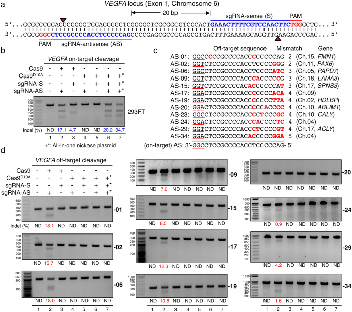
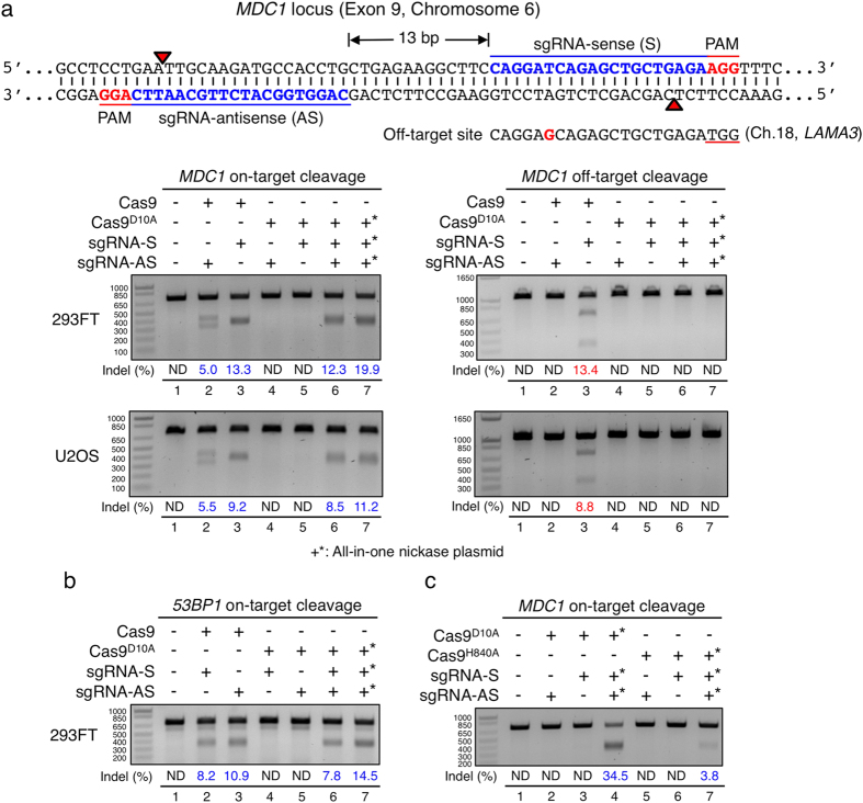
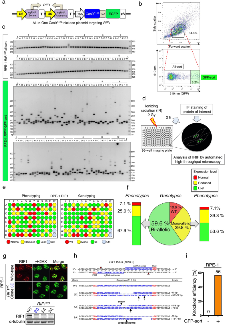
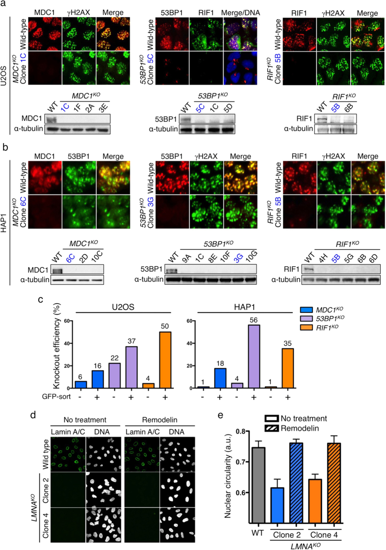
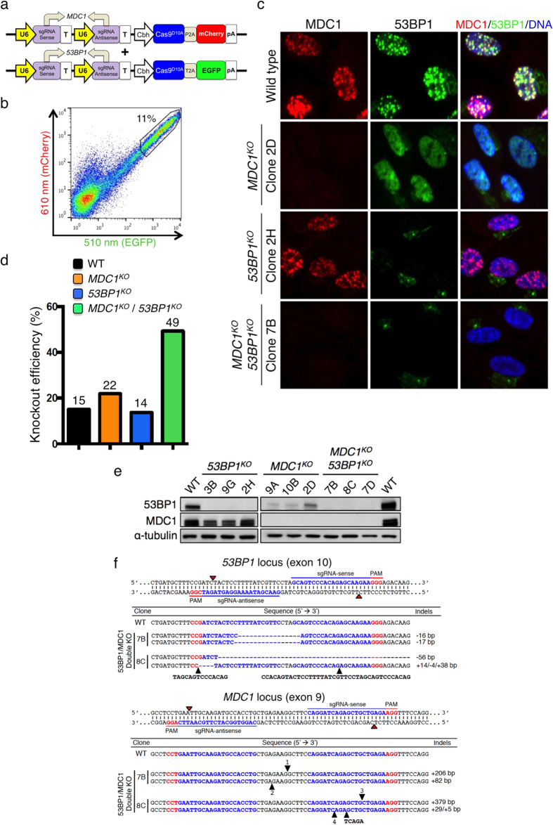
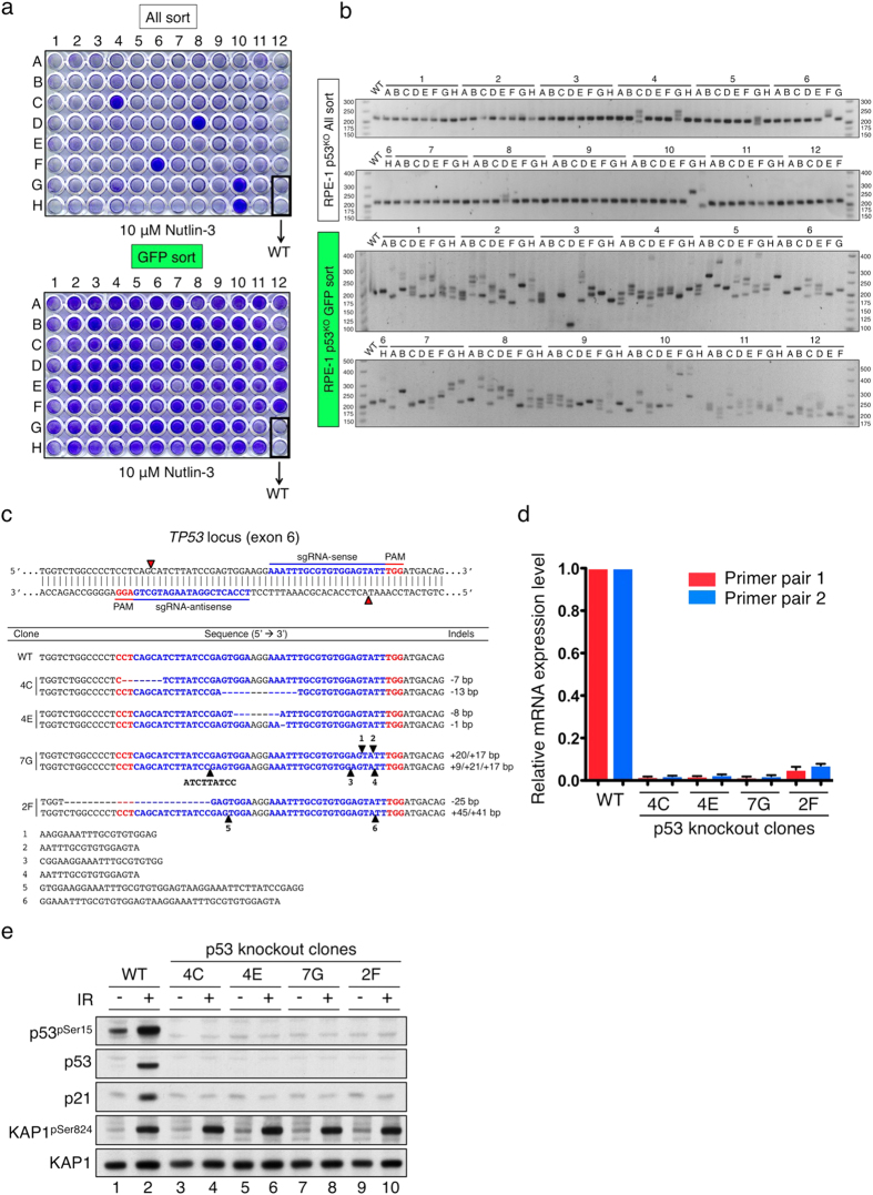
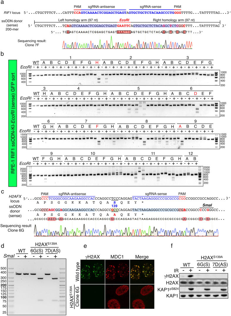
The RNA-guided Cas9 nuclease is being widely employed to engineer the genomes of various cells and organisms. Despite the efficient mutagenesis induced by Cas9, off-target effects have raised concerns over the system's specificity. Recently a "double-nicking" strategy using catalytic mutant Cas9(D10A) nickase has been developed to minimise off-target effects. Here, we describe a Cas9(D10A)-based screening approach that combines an All-in-One Cas9(D10A) nickase vector with fluorescence-activated cell sorting enrichment followed by high-throughput genotypic and phenotypic clonal screening strategies to generate isogenic knockouts and knock-ins highly efficiently, with minimal off-target effects. We validated this approach by targeting genes for the DNA-damage response (DDR) proteins MDC1, 53BP1, RIF1 and P53, plus the nuclear architecture proteins Lamin A/C, in three different human cell lines. We also efficiently obtained biallelic knock-in clones, using single-stranded oligodeoxynucleotides as homologous templates, for insertion of an EcoRI recognition site at the RIF1 locus and introduction of a point mutation at the histone H2AFX locus to abolish assembly of DDR factors at sites of DNA double-strand breaks. This versatile screening approach should facilitate research aimed at defining gene functions, modelling of cancers and other diseases underpinned by genetic factors, and exploring new therapeutic opportunities.


Figures

Figure 1
Figure 1. On- and off-target mutagenic efficiencies of sgRNAs targeting VEGFA in human cells.
(a) Double nicking strategy with sense (S) and antisense (AS) sgRNAs separated by 20 base pairs at exon 1 of the VEGFA locus on chromosome 6. Red arrows indicate the nicking sites of Cas9D10A nickase, which generates 5′ overhangs at target sites. (b) On-target mutagenic efficiencies of VEGFA sgRNAs by T7 EI assays in human HEK293FT cells. Cells were either transfected with or without (lane 1) wild-type Cas9 vector containing either the AS (lane 2) or the S sgRNA (lane 3), single sgRNA Cas9D10A nickase vector carrying either the AS (lane 4) or the S sgRNA (lane 5), two single sgRNA nickase vectors each containing Cas9D10A nickase with either the S or the AS sgRNA (lane 6), or dual sgRNA All-in-One Cas9D10A nickase vector (lane 7). Indel frequencies are shown below. ND, not detected. (c) Twelve off-target sites of the AS sgRNA were assessed. Mismatch nucleotides are indicated in red, and PAM motifs (NGG) are underlined. The number of mismatches, associated genes and chromosomes are indicated. (d) Off-target cleavages by wild-type Cas9 with the AS sgRNA at eleven off-target sites assessed. Note that we were unable to get conclusive results at AS-23 site where PCR products, amplified by two different sets of primers, were unexpectedly degraded by T7 EI across all samples, which may arise from high heterogeneity at this particular genomic locus. +*All-in-One nickase plasmid.
Figure 2
Figure 2. On- and off-target mutagenic efficiencies of sgRNAs targeting MDC1 or 53BP1.
(a) Double nicking strategy with sense (S) and antisense (AS) sgRNAs separated by 13 base pairs at exon 9 of the MDC1 locus on chromosome 6. One verified off-target site of the S sgRNA (located on chromosome 18, LAMA3) is shown below with one mismatch indicated in red. The on- and off-target mutagenic efficiencies were assessed by T7 EI assays in human HEK293FT and U2OS cells. Cells were either transfected with or without (lane 1) wild-type Cas9 vector carrying either the AS (lane 2) or the S sgRNA (lane 3), single-sgRNA Cas9D10A nickase vector containing either the AS (lane 4) or the S sgRNA (lane 5), two single sgRNA nickase vectors each carrying Cas9D10A nickase with either the S or the AS sgRNA (lane 6), or dual sgRNA All-in-One Cas9D10A nickase vector (lane 7). (b) On-target mutagenic efficiencies of sgRNAs targeting the exon 10 of 53BP1 locus. (c) On-target mutagenic efficiencies by Cas9D10A or Cas9H840A nickases at the MDC1 locus. Cells were either transfected with or without (lane 1) single sgRNA Cas9D10A nickase vector containing either the AS (lane 2) or the S sgRNA (lane 3), All-in-One Cas9D10A nickase vector (lane 4), single sgRNA Cas9H840A nickase vector carrying either the AS sgRNA (lane 5) or the S sgRNA (lane 6), or All-in-One Cas9H840A nickase vector (lane 7). +*All-in-One nickase plasmid.
Figure 3
Figure 3. Genotypic and phenotypic screens for RIF1 knockout clones in RPE-1 cells.
(a) Schematic of the All-in-One Cas9D10A nickase vector targeting RIF1. (b) FACS data showing the sequential gating of healthy (all-sort, 64.4%, top panel) and high-EGFP expressing (GFP-sort, 6.2%, bottom panel) cell populations. Each of these populations was sorted separately into 96-well plates at the level of a single cell per well. (c) PCR-genotyping of 94 clones from all- and GFP-sorted populations to identify indels at the target site. Wild-type (WT) product size is 182 bp. (d) Schematic of the automated high-throughput microscopy-based phenotypic screen for RIF1 knockout clones. Cells were subjected to 2 Gy of IR. After 2 h, immunofluorescent cell staining was carried out for RIF1 protein. IR-induced foci were then analysed by high-throughput automatic microscopy. (e,f) Genotypic and phenotypic screening outputs of RIF1 knockout clones were compared in parallel. In genotyping panel: WT, clones with no apparent indel formation. Mono, clones with monoallelic indel mutations. Bi, clones with biallelic indel mutations. Ctrl, wild-type non-targeted clones. In phenotyping panel: Normal, normal foci equivalent to wild-type. Reduced, reduced foci compared to wild-type. Lost, clones with no staining of foci. (g) Representative IF images of RPE-1 RIF1 knockout and wild-type cells from high-throughput microscopy screening. Cells were co-stained with RIF1 and γH2AX antibodies. Level of RIF1 protein was verified by Western blotting, shown below. (h) DNA sequencing of RIF1 knockout clones at both alleles. Red arrowheads indicate the Cas9D10A nicking sites. Each sequence represents one allele. Dashed lines indicate deletions; black arrowheads indicate the precise locations of insertions. Inserted sequences are either tagged with black arrowheads, or numbered and shown in Supplementary Fig. 3. (i) Phenotypic RIF1 knockout efficiencies of all-sort and GFP-sort clones in RPE-1 cells.
Figure 4
Figure 4. High-throughput microscopy-based phenotypic screening for knockout clones in human U2OS and HAP1 cells.
(a) Representative IF images of knockout clones for MDC1, 53BP1 and RIF1 in U2OS cells from the automated high-throughput microscopy screen. In the middle panel, merge/DNA indicates the overlay of 53BP1, RIF1 and DAPI (DNA) staining. Western blots are shown below. (b) Representative IF images of knockout clones for MDC1, 53BP1 and RIF1 in HAP1 cells. (c) Phenotypic knockout efficiencies of all-sort and GFP-sort clones in U2OS and HAP1 cells. (d) Confocal microscopy images of LMNA knockouts in U2OS cells with or without Remodelin treatment. Cells were co-stained with Lamin A/C antibody and DAPI (DNA). (e) Quantification of nuclear circularities of wild-type and LMNA knockout cells in the presence or absence of Remodelin.
Figure 5
Figure 5. Simultaneous double knockout of MDC1 and 53BP1.
(a) Sense and antisense sgRNAs targeting MDC1 and 53BP1 were cloned into 2A-linked mCherry and EGFP All-in-One Cas9D10A nickase vectors, respectively. (b) FACS single-cell sorting of cell populations co-expressing mCherry and EGFP gated for the top 11%. (c) Representative high-throughput microscopy IF images of wild-type, MDC1 knockout, 53BP1 knockout, and MDC1 and 53BP1 double knockout (MDC1KO/53BP1KO) RPE-1 cells. 53BP1/MDC1/DNA indicates the overlay of 53BP1, MDC1 and DAPI (DNA) staining. (d) Phenotypic knockout efficiencies of respective knockout populations. (e) Western immunoblots of respective knockout clones. (f) DNA sequencing of two MDC1KO/53BP1KO clones across the MDC1 and 53BP1 targeting loci. The full sequences of numbered insertions are shown in Supplementary Fig. 11f.
Figure 6
Figure 6. Drug-based (Nutlin-3) functional screen for TP53 knockouts.
(a) Nutlin-3 drug screen to identify p53 knockouts from all- and GFP-sort clones that were plated at a similar density and treated with 10 μM Nutlin-3 for five days prior to fixation and crystal violet staining. (b) PCR-genotyping of 94 clones from all- and GFP-sort populations to identify indels at the TP53 target site. Wild-type (WT) product size is 221 bp. (c) DNA sequencing of TP53 knockout clones, confirming biallelic disruption. (d) qRT-PCR of TP53 mRNA levels in wild-type and knockout clones. (e) Functional validation of four TP53 knockout clones with regard to DDR signalling. Cells were damaged with or without 10 Gy of IR. 8 h later, cells were harvested and analysed by Western immunoblotting. KAP1pS824 is a marker of DNA damage.
Figure 7
Figure 7. Precise biallelic knock-ins via HDR by Cas9D10A nickase-based system.
(a) Strategic knock-in insertion of an EcoRI site at exon 3 of the RIF1 locus. Only the coding strand is shown. The intended mutations in the sense ssODN template are indicated in red. DNA sequencing of a homozygous knock-in clone 7F is aligned below. (b) Genotyping analysis of the PCR products across the knock-in site by EcoRI digestion. Clones indicated in red were verified by DNA sequencing as biallelic knock-ins where EcoRI-digested products were 487 bp and 310 bp. (c) H2AXS139A point mutation knock-in strategy. Only the coding strand is shown and the corresponding amino acid sequence is aligned below. In the ssODN template, eight nucleotide mutations, including creation of a SmaI site, are indicated in red. DNA sequencing for homozygous knock-in clone 6G is aligned at the bottom. Successful knock-in nucleotides are indicated in red boxes. (d) Genotyping analysis of the PCR products across the H2AFX target site by SmaI digestion. Undigested product length is 363 bp and SmaI-digested products were 287 bp and 76 bp. (e) IRIF of wild-type and H2AXS139A cells. Cells were subjected to 2 Gy of IR and fixed after 2 h, followed by γH2AX and MDC1 immunostaining. (f) Western immunoblots of wild-type and H2AXS139A clones subjected to 10 Gy of IR and harvested after 20 min. KAP1pS824 is a marker of DNA damage.

References

    1. Jinek M. et al. A programmable dual-RNA-guided DNA endonuclease in adaptive bacterial immunity. Science 337, 816–821, doi: 10.1126/science.1225829 (2012). - DOI - PMC - PubMed
    1. Garneau J. E. et al. The CRISPR/Cas bacterial immune system cleaves bacteriophage and plasmid DNA. Nature 468, 67–71, doi: 10.1038/nature09523 (2010). - DOI - PubMed
    1. Wiedenheft B., Sternberg S. H. & Doudna J. A. RNA-guided genetic silencing systems in bacteria and archaea. Nature 482, 331–338, doi: 10.1038/nature10886 (2012). - DOI - PubMed
    1. Horvath P. & Barrangou R. CRISPR/Cas, the immune system of bacteria and archaea. Science 327, 167–170, doi: 10.1126/science.1179555 (2010). - DOI - PubMed
    1. Ran F. A. et al. Genome engineering using the CRISPR-Cas9 system. Nat. Protoc. 8, 2281–2308, doi: 10.1038/nprot.2013.143 (2013). - DOI - PMC - PubMed

Publication types

MeSH terms

Substances