. 2016 Jun;36(6):1209-19.
doi: 10.1161/ATVBAHA.116.307517. Epub 2016 Apr 14.

An Intronic Flk1 Enhancer Directs Arterial-Specific Expression via RBPJ-Mediated Venous Repression

Affiliations

An Intronic Flk1 Enhancer Directs Arterial-Specific Expression via RBPJ-Mediated Venous Repression

Philipp W Becker et al. Arterioscler Thromb Vasc Biol. 2016 Jun.

Abstract

Objective: The vascular endothelial growth factor (VEGF) receptor Flk1 is essential for vascular development, but the signaling and transcriptional pathways by which its expression is regulated in endothelial cells remain unclear. Although previous studies have identified 2 Flk1 regulatory enhancers, these are dispensable for Flk1 expression, indicating that additional enhancers contribute to Flk1 regulation in endothelial cells. In the present study, we sought to identify Flk1 enhancers contributing to expression in endothelial cells.

Approach and results: A region of the 10th intron of the Flk1 gene (Flk1in10) was identified as a putative enhancer and tested in mouse and zebrafish transgenic models. This region robustly directed reporter gene expression in arterial endothelial cells. Using a combination of targeted mutagenesis of transcription factor-binding sites and gene silencing of transcription factors, we found that Gata and Ets factors are required for Flk1in10 enhancer activity in all endothelial cells. Furthermore, we showed that activity of the Flk1in10 enhancer is restricted to arteries through repression of gene expression in venous endothelial cells by the Notch pathway transcriptional regulator Rbpj.

Conclusions: This study demonstrates a novel mechanism of arterial-venous identity acquisition, indicates a direct link between the Notch and VEGF signaling pathways, and illustrates how cis-regulatory diversity permits differential expression outcomes from a limited repertoire of transcriptional regulators.

Keywords: arterial-venous specification; artery; endothelial cells; mice; notch; veins; zebrafish.


Figures

Figure 1.
Figure 1.
The mouse Flk1in10 enhancer directs arterial-restricted expression in transgenic mice. A, Schematic representation of the mouse Flk1in10 enhancer (top line, exons are black boxes) and Flk1in10:LacZ transgene (bottom line). B–K, The Flk1in10:LacZ transgene directs arterial expression. Representative whole-mount embryos and yolk sac tissue from the Flk1in10:LacZ transgenic line (B–F) show reporter gene expression (X-gal staining, blue) in the vasculature from embryonic day 8 (E8) to E16. X-gal staining is initially detected throughout the vasculature but becomes restricted to the arterial compartment during development. G–J, In transverse sections through E9–E12 transgenic embryos, X-gal staining is detected in both cardinal vein and dorsal aorta at E9 (G) but is stronger in the dorsal aorta by E10 and is not detected in the venous or lymphatic vasculature by E12. K, E12 transverse paraffin sections showed that expression of the venous marker endomucin does not overlap with that of the β-galactosidase reporter gene. a indicates artery; al, allantois; ca, carotid artery; cv, cardinal vein; da, dorsal aorta; jls, jugular lymph sac; jv, jugular vein; np, neural plexus; and v, vein.
Figure 2.
Figure 2.
The mouse Flk1in10 enhancer directs arterial-restricted expression in transgenic zebrafish. A, Schematic representation of the Flk1in10:GFP transgene. B, The mouse Flk1in10:GFP transgene directs endothelial cell–specific expression in transgenic zebrafish line tg(Flk1in10:GFP) at 24 h post fertilization (hpf). C–E, Tg(Flk1in10:GFP;kdrl:HRAS:mCherry) zebrafish expresses GFP in most endothelial cells at 30 hpf (C). At 48 hpf, GFP expression in the axial vessels becomes restricted to the dorsal aorta and to a subset of intersegmental vessels, corresponding to the developing intersegmental arteries (ISA; D; see also Movie I in the online-only Data Supplement). At 72 hpf, little dorsal aorta expression could be detected, while GFP expression is maintained in the ISA and dorsal longitudinal anastomotic vessel (E). F, Bar chart detailing GFP expression pattern in the ISA and intersegmental veins (ISVe) at 72 hpf. Represents a total of 18 embryos, ISA and ISVe identity established by using kdrl:HRAS-mCherry expression to determine whether each vessel connected to dorsal aorta or posterior cardinal vein. Any detectable level of GFP expression constituted positive, and Figure III in the online-only Data Supplement details analysis methods. bc indicates blood cells; da, dorsal aorta; dlav, dorsal longitudinal anastomotic vessel; dv, dorsal vein; GFP, green fluorescent protein; isv, intersegmental vessels; ISVe, intersegmental vein; and vv, ventral vein.
Figure 3.
Figure 3.
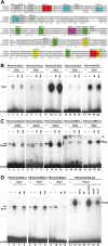
The Flk1in10 enhancer contains cis-motifs that bind Etv2, Gata2, Rbpj, Sox7, and Foxc2 transcription factors. A, Multispecies alignment of the conserved region of the Flk1in10 enhancer. Colored sequences depict confirmed consensus binding motifs, and gray sequences depict motifs that did not bind in electrophoretic mobility shift assay. B, Radiolabeled oligonucleotide probes encompassing Flk1in10 ETS-f (lanes 1–4), ETS-g (lanes 5–8), ETS-h (lanes 9–12), ETS-k (lanes 13–16), and ETS-l motifs (lanes 17–20) were bound to recombinant Etv2 proteins. All proteins efficiently bound to labeled probes (lanes 2, 6, 10, 14, and 18) were competed by excess unlabeled self-probe (wt, lanes 3, 7, 11, 15, and 19) but not by mutant self-probe (mu, lanes 4, 8, 12, 16, and 20). C, Radiolabeled oligonucleotide probes encompassing Flk1in10 GATA-b (lanes 1–4), GATA-c (lanes 5–8), GATA-d (lanes 9–12), RBPJ-a (lanes 13–16), and RBPJ-b motifs (lanes 17–20) were bound to recombinant Gata2 and Rbpj proteins. All proteins efficiently bound to labeled probes (lanes 2, 6, 10, 14, and 18) were competed by excess unlabeled self-probe (wt, lanes 3, 7, 11, 15, and 19) but not by mutant self-probe (mu, lanes 4, 8, 12, 16, and 20). D, Radiolabeled oligonucleotide probes encompassing Flk1in10 SOX-a (lanes 1–4), SOX-b (lanes 5–8), SOX-c (lanes 9–12), and FOX-a/b (lanes 13–19) were bound to recombinant Sox7 and Foxc2 proteins. All proteins efficiently bound to labeled probes (lanes 2, 6, 10, and 14) were competed by excess unlabeled self-probe (wt, lanes 3, 7, 11, and 16) but not by mutant self-probe (mu, lanes 4, 8, and 12). For FOX-a/b, mutant self-probe with mutations to both FOX motifs (mu A+B) or to FOX-a could not compete with labeled probes (17, 19), whereas a mutant self-probe with mutations to FOX-b (mu B) was still able to robustly compete with labeled probe (18). mu indicates mutant; ns, nonspecific binding; and wt, wild-type.
Figure 4.
Figure 4.
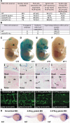
The Flk1in10 enhancer requires ETS and GATA motifs for endothelial expression. A and B, Summary of reporter gene expression detected in (A) 48 hpf Tol2-mediated mosaic transient transgenic zebrafish embryos and (B) E12 transient transgenic mice. Asterisk indicates weak staining in head region only. C–F, Whole-mount (top) and transverse sections (bottom) of representative E12 X-gal–stained embryos transgenic for the Flk1in10mutGATA-b,c construct. Depicted sections correspond to the embryos shown. Number in bottom corner of whole-mount embryo denotes the number of embryos similar to that shown. Lines marked i and ii on C mark the approximate location of transverse sections. Arrowheads mark head vessels. G, Analysis of scrambled, 1.125 ng, 2.25 ng, and 3.375 ng gata2a morpholino (MO) in 48 h post fertilization (hpf) tg(Flk1in10:GFP) embryos. H, Analysis of scrambled, 2.25 ng and 3.375 ng gata2a MO in 26 hpf WT zebrafish embryos, using whole-mount kdr in situ hybridization. cv indicates cardinal vein; da, doral aorta; and GFP, green fluorescent protein.
Figure 5.
Figure 5.
Rbpj is required for arterial restriction of the Flk1in10 enhancer. A and B, Summary of reporter gene expression detected in (A) 48 h post fertilization (hpf) Tol2-mediated mosaic transient transgenic zebrafish embryos and (B) embryonic day (E) 12 transient transgenic mice. C–F, Whole-mount (top) and transverse sections (bottom) of representative E12 X-gal–stained transient transgenic embryos expressing mouse Flk1in10 WT (C), mouse Flk1in10mRBPJ/SOX (D), mouse Flk1in10mRBPJ (E), and chicken Flk1in10 WT (F). Lines marked i and ii on C mark the approximate location of transverse sections in C–F. a indicates artery; cv, cardinal vein; da, dorsal aorta; and v, veins. G–I, Analysis of the effects of scrambled (G), 1.25 ng rbpj morpholino (MO) oligonucleotides (H), and Notch signaling inhibitor N-[N-3,5-difluorophenacetyl]-l-alanyl-S-phenylglycine methyl ester (DAPM) (I) in 72 h post fertilization (hpf) tg(Flk1in10:GFP) embryos. MO-mediated knockdown of rbpj results in expansion of Flk1in10:GFP expression into the caudal vein, the posterior cardinal vein, and intersegmental vein. da indicates dorsal aorta; GFP, green fluorescent protein; ISA, intersegmental artery; ISVe, intersegmental vein; and pcv, posterior cardinal vein.
Figure 6.
Figure 6.
Ablation of RBPJ and SOX motifs alongside perturbation of GATA sites reveals a requirement for Rbpj/Notch and SoxF factors in the activation of Flk1in10 in arteries. A and B, Summary of reporter gene expression detected in (A) 48 h post fertilization (hpf) Tol2-mediated mosaic transient transgenic zebrafish embryos and (B) embryonic day (E) 12 transient transgenic mice. C and D, Bar charts representing zebrafish (C) and mouse (D) expression patterns of Flk1in10 constructs reported in A and B. E, Whole-mount embryos (top) and yolk sac tissues (bottom) of representative E12 X-gal–stained transient transgenic embryos expressing mouse Flk1in10mutGATA-a,b,d, mouse Flk1in10mutGATA-a,b,d/RBPJ/SOX, mouse Flk1in10mutGATA-b, and mouse Flk1in10mutGATA-b/altRBPJ/altSOX (E). a indicates artery; GFP, green fluorescent protein; and v, veins.

References

    1. Jin SW, Patterson C. The opening act: vasculogenesis and the origins of circulation. Arterioscler Thromb Vasc Biol. 2009;29:623–629. doi: 10.1161/ATVBAHA.107.161539. - PMC - PubMed
    1. Potente M, Gerhardt H, Carmeliet P. Basic and therapeutic aspects of angiogenesis. Cell. 2011;146:873–887. doi: 10.1016/j.cell.2011.08.039. - PubMed
    1. Lawson ND, Scheer N, Pham VN, Kim CH, Chitnis AB, Campos-Ortega JA, Weinstein BM. Notch signaling is required for arterial-venous differentiation during embryonic vascular development. Development. 2001;128:3675–3683. - PubMed
    1. Sacilotto N, Monteiro R, Fritzsche M, Becker PW, Sanchez-Del-Campo L, Liu K, Pinheiro P, Ratnayaka I, Davies B, Goding CR, Patient R, Bou-Gharios G, De Val S. Analysis of Dll4 regulation reveals a combinatorial role for Sox and Notch in arterial development. Proc Natl Acad Sci USA. 2013;110:11893–11898. doi: 10.1073/pnas.1300805110. - PMC - PubMed
    1. You LR, Lin FJ, Lee CT, DeMayo FJ, Tsai MJ, Tsai SY. Suppression of Notch signalling by the COUP-TFII transcription factor regulates vein identity. Nature. 2005;435:98–104. doi: 10.1038/nature03511. - PubMed

MeSH terms

Substances