. 2016 Apr 15:6:24454.
doi: 10.1038/srep24454.

Computer-Aided Diagnosis with Deep Learning Architecture: Applications to Breast Lesions in US Images and Pulmonary Nodules in CT Scans

Affiliations

Computer-Aided Diagnosis with Deep Learning Architecture: Applications to Breast Lesions in US Images and Pulmonary Nodules in CT Scans

Jie-Zhi Cheng et al. Sci Rep. .

Abstract

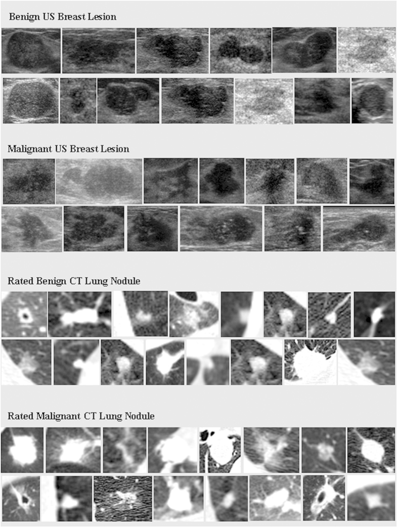
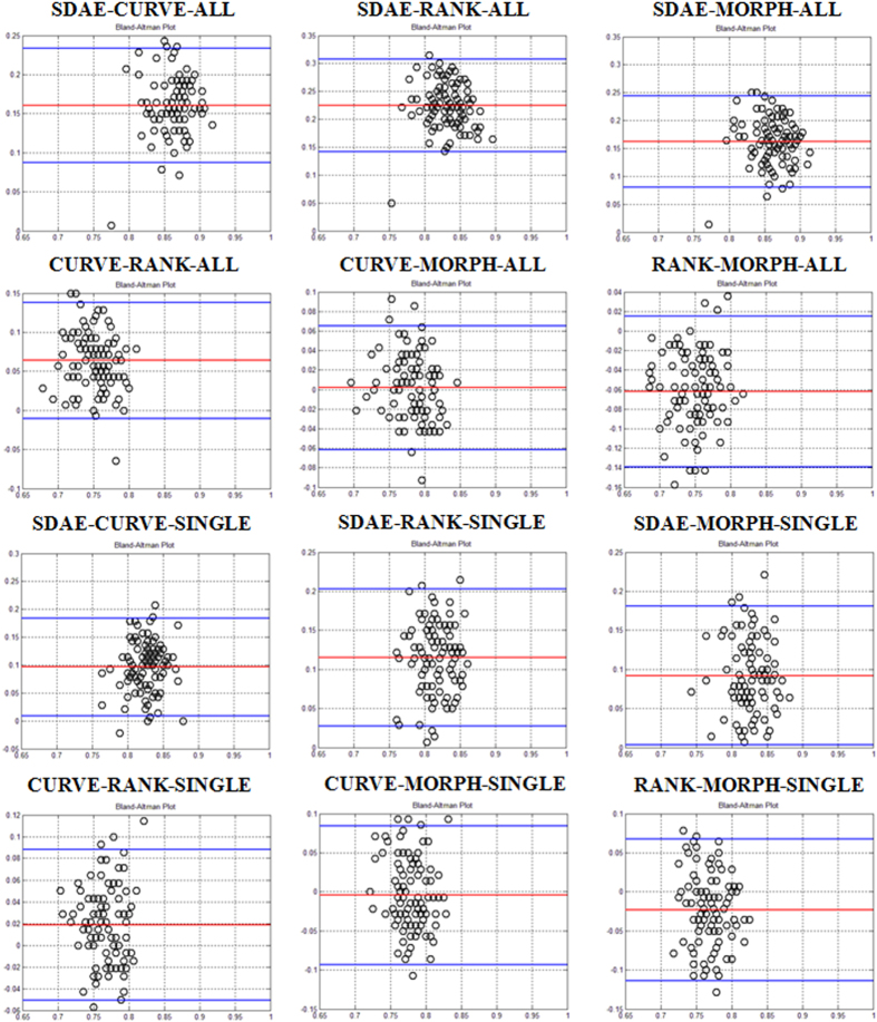
This paper performs a comprehensive study on the deep-learning-based computer-aided diagnosis (CADx) for the differential diagnosis of benign and malignant nodules/lesions by avoiding the potential errors caused by inaccurate image processing results (e.g., boundary segmentation), as well as the classification bias resulting from a less robust feature set, as involved in most conventional CADx algorithms. Specifically, the stacked denoising auto-encoder (SDAE) is exploited on the two CADx applications for the differentiation of breast ultrasound lesions and lung CT nodules. The SDAE architecture is well equipped with the automatic feature exploration mechanism and noise tolerance advantage, and hence may be suitable to deal with the intrinsically noisy property of medical image data from various imaging modalities. To show the outperformance of SDAE-based CADx over the conventional scheme, two latest conventional CADx algorithms are implemented for comparison. 10 times of 10-fold cross-validations are conducted to illustrate the efficacy of the SDAE-based CADx algorithm. The experimental results show the significant performance boost by the SDAE-based CADx algorithm over the two conventional methods, suggesting that deep learning techniques can potentially change the design paradigm of the CADx systems without the need of explicit design and selection of problem-oriented features.


Figures

Figure 1
Figure 1. Exhibition of breast lesions and lung nodules in US and CT images.
Figure 2
Figure 2
Examples of constructed patterns in the first and second hidden layers at the pre-training step: (a,b) patterns of the first and second hidden layers for pulmonary nodules; (c,d) patterns of the first and second hidden layers for breast lesions. SDAE architecture with two hidden layers is used in this study for the differentiation of pulmonary nodules and breast lesions. It is worth noting that the patterns of the second hidden layers are constructed as the weighted sums from all patterns in the first layer. In the reconstruction, the first layer neurons are simply all assumed activated. The neuron activation can be more complicated with the feed-in of real image data. In (b,d) the example patterns enclosed by the yellow rectangles hold the positive weightings to the RN nodule and benignant lesion classes in the supervised training step, whereas the patterns in blue regions are connected to the RM nodule and malignant lesion classes with positive weightings. It can be observed from (b,d) that the second hidden layer patterns appear fuzzier due to the effect of weighted sum. All patterns are normalized for clearer presentation.
Figure 3
Figure 3. ACC Bland and Altman plots for six algorithm comparing pairs of “SDAE-CURVE”, “SDAE-RANK” , “SDAE-MORPH”, “CURVE-RANK”, “CURVE-MORPH”, and “RANK-MORPH” on the lung CT dataset.
The comparing pairs with ending tag “ALL” are the results with the strategy of using all member slices of a nodule for the training and testing of the three algorithms. The pairs with tag “SINGLE” compare the computerized results with the slice selection strategy of using middle slice.
Figure 4
Figure 4. ACC Bland and Altman plots for performance comparison of the pairs “SDAE-CURVE”, “SDAE-RANK”, “SDAE-MORPH”, “CURVE-RANK”, “CURVE-MORPH”, and “RANK-MORPH” on the breast dataset.
Figure 5
Figure 5. Box plots for performance for the lung and breast datasets with respect to the ACC and AUC metrics.
Figure 6
Figure 6. Flow-chart of our deep-learning-based CADx training framework.
The pixels of resized ROIs are fed into the network architecture at the pre-training step. The pre-trained network is then refined with the supervised training by adding three neurons carrying aspect ratio of the original ROI and also the resizing factors at the input layer. The final identification result can be made with the softmax classification.

References

    1. Doi K. Computer-aided diagnosis in medical imaging: historical review, current status and future potential. Comput. Med. Imaging Graph. 31, 198–211 (2007). - PMC - PubMed
    1. van Ginneken B., Schaefer-Prokop C. M. & Prokop M. Computer-aided diagnosis: how to move from the laboratory to the clinic. Radiology 261, 719–732 (2011). - PubMed
    1. Giger M. L., Chan H.-P. & Boone J. Anniversary paper: history and status of CAD and quantitative image analysis: the role of medical physics and AAPM. Med. Phys. 35, 5799–5820 (2008). - PMC - PubMed
    1. Cheng J.-Z. et al. Computer-aided US diagnosis of breast lesions by using cell-based contour grouping1. Radiology 255, 746–754 (2010). - PubMed
    1. Giger M. L., Karssemeijer N. & Schnabel J. A. Breast image analysis for risk assessment, detection, diagnosis, and treatment of cancer. Annu. Rev. Biomed. Eng. 15, 327–357 (2013). - PubMed

Publication types