. 2016 May;23(5):142-8.
doi: 10.1038/cgt.2016.14. Epub 2016 Apr 15.

Regional CAR-T cell infusions for peritoneal carcinomatosis are superior to systemic delivery

Affiliations

Regional CAR-T cell infusions for peritoneal carcinomatosis are superior to systemic delivery

S C Katz et al. Cancer Gene Ther. 2016 May.

Abstract

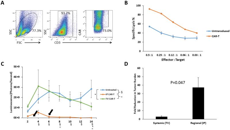
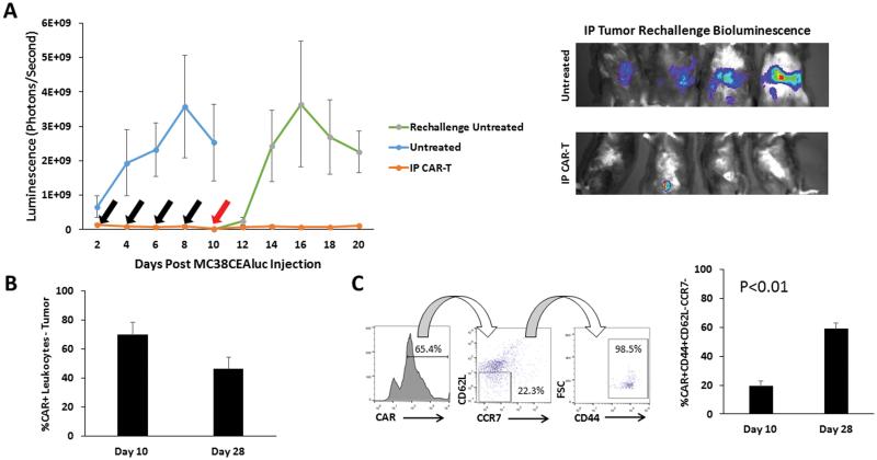
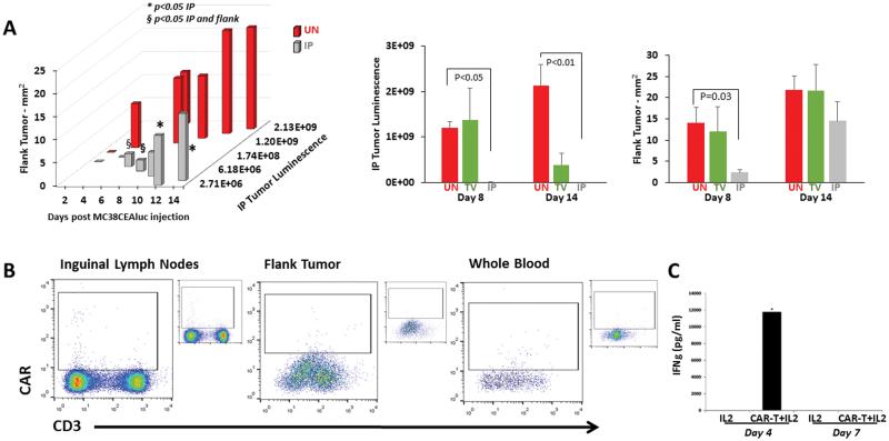
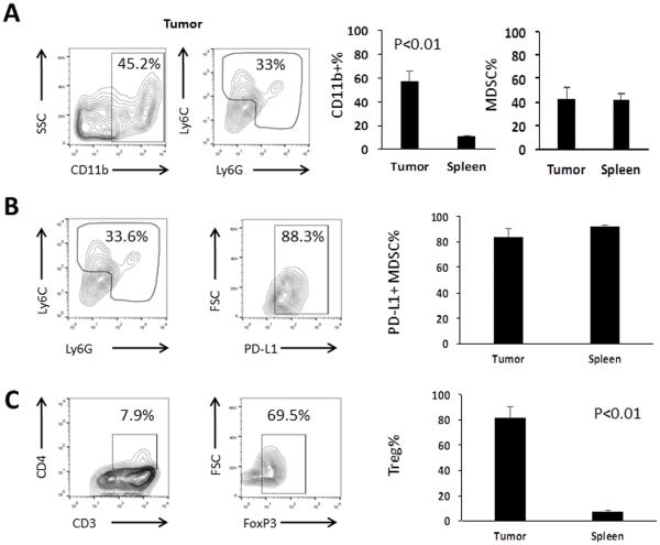
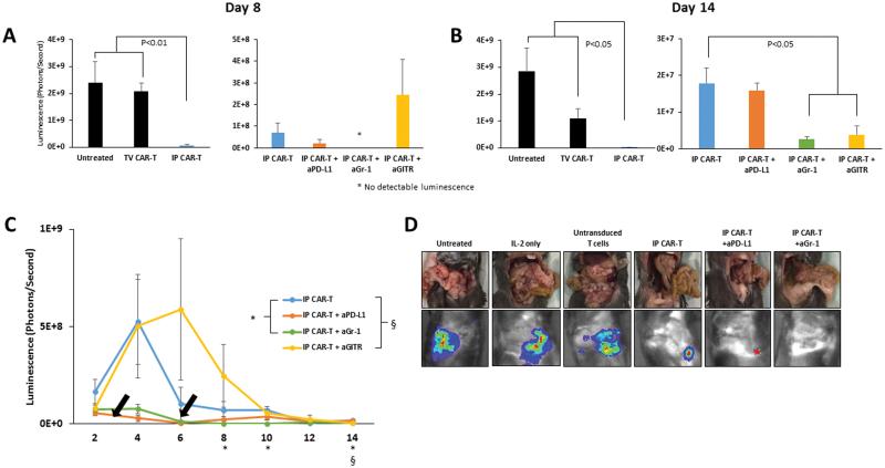
Metastatic spread of colorectal cancer (CRC) to the peritoneal cavity is common and difficult to treat, with many patients dying from malignant bowel obstruction. Chimeric antigen receptor T cell (CAR-T) immunotherapy has shown great promise, and we previously reported murine and phase I clinical studies on regional intrahepatic CAR-T infusion for CRC liver metastases. We are now studying intraperitoneal (IP) delivery of CAR-Ts for peritoneal carcinomatosis. Regional IP infusion of CAR-T resulted in superior protection against carcinoembryonic antigen (CEA+) peritoneal tumors, when compared with systemically infused CAR-Ts. IP CAR-Ts also provided prolonged protection against IP tumor re-challenges and demonstrated an increase in effector memory phenotype over time. IP CAR-Ts provided protection against tumor growth at distant subcutaneous (SC) sites in association with increases in serum IFNγ levels. Given the challenges posed by immunoinhibitory pathways in solid tumors, we combined IP CAR-T treatment with suppressor cell targeting. High frequencies of myeloid-derived suppressor cells (MDSC) and regulatory T cells (Treg) were found within the IP tumors, with MDSC expressing high levels of immunosuppressive PD-L1. Combinatorial IP CAR-T treatment with depleting antibodies against MDSC and Treg further improved efficacy against peritoneal metastases. Our data support further development of combinatorial IP CAR-T immunotherapy for peritoneal malignancies.


Figures

Figure 1
Figure 1. Optimization of CAR-T delivery for treatment of peritoneal tumors
CAR-Ts were generated from murine splenocytes and were characterized for expression of CD3 and the anti-CEA CAR (A). In vitro CAR-T tumor killing was tested with bioluminescence assays, in which CAR-Ts or untransduced T cells were co-cultured with MC38CEA-luc at various effector : target ratios (B). CAR-Ts were tested in vivo via IP or TV infusion, and peritoneal tumor killing was monitored by changes in bioluminescence. Arrows indicate the CAR-T injection time points. Each line on the plot is representative of the average of 4 mice. Symbols denoting significance (p<0.05) are found below the x-axis (C). Fold reduction in tumor luminescence was calculated between days 4 and 14 of the in vivo study, comparing TV to IP CAR-T delivery (D). Error bars are representative of SEM values. P values were calculated using Student’s t test.
Figure 2
Figure 2. Durability of IP CAR-T tumor efficacy in mice with peritoneal tumors
Duration of CAR-T anti-tumor efficacy in the peritoneal space was tested with IP tumor re-challenge, following response to initial tumor burden. Black arrows represent days that mice received CAR-T treatment and the red arrow represents the day of tumor re-challenge (A, left). Tumor growth was measured by bioluminescence. Representative bioluminescence images from day 20 of the study are depicted from mice that received tumor re-challenge (A, right). The frequencies of CAR+ lymphocytes recovered from IP tumor tissue at both day 10 (n=5) and day 28 (n=3) time points were compared (B). Memory phenotypes of CAR+ lymphocytes were examined at both the day 10 (n=5) and day 28 (n=3) time points. Dot plots show the gating for the effector memory phenotype (C). Error bars are representative of SEM values. P values were calculated using Student’s t test.
Figure 3
Figure 3. Systemic response to IP CAR-Ts
The systemic anti-tumor effect of regionally delivered IP CAR-Ts was tested by injecting mice with MC38CEA-luc both IP and subcutaneously in the left flank. IP tumor burden and flank tumor sizes were measured for 14 days in untreated (UN), TV CAR-T and IP CAR-T groups. Bars are representative of 4 mice per group. (A). On day 14, left inguinal lymph nodes, flank tumors, and whole blood were harvested to determine if CAR-Ts were present at these sites. Fully stained, untreated controls are shown in insets next to the dot plots (B). Error bars are representative of SEM values. P values were calculated using Student’s t test. Bioluminescence is expressed as photons per second. (C) Serum IFNγ concentrations were determined on days 4 and 7 to detect systemic immune activity in response to IP CAR-T infusions.
Figure 4
Figure 4. Suppressor cell content of intraperitoneal tumors
Tumor leukocyte contents were immunophenotyped to detect the presence of suppressive cell populations. MDSC were found in the tumors after staining for CD11b, Ly6C and Ly6G. Representative dot plots show MDSC from the IP tumors, along with bar graphs comparing MDSC populations from the tumors and spleens of the same untreated animals. The percentages of CD11b+ cells among all live cells and MDSC (Gr-1+) among CD11b+ cells are shown (A). MDSC were also immunophenotyped for the expression of the immunosuppressive marker PD-L1 (B). Representative tumor dot plots show that Treg, expressed as the percentage of FoxP3+ cells among CD3+CD4+ T cells, were also found within the IP tumors. Smaller populations were found within the spleens of the same animals (C). Bars are representative of 3 mice per group. Error bars are representative of SEM values. P values were calculated using Student’s t test.
Figure 5
Figure 5. IP CAR-Ts in combination with suppressor cell targeting for in vivo treatment
IP CAR-Ts combined with depleting antibodies against MDSC and Treg, or blocking antibodies against the PD-L1 pathway, were administered to mice injected with MC38CEA-luc. Tumor reduction was monitored by bioluminescence over 14 days. Bar graphs compare the efficacy of regional IP CAR-Ts to systemic TV CAR-Ts, and IP CAR-Ts alone to IP CAR-Ts with antibodies on day 8 after the treatments (A) and at the end of the study on day 14 (B). Bars are representative of 4 animals per group. Black arrows represent CAR-T treatments. Symbols denoting significance appear for specific days below the x axis (C). Gross inspection images, as well as bioluminescence images, are shown for all groups on day 14 (D). Error bars are representative of SEM values. P values were calculated using Student’s t test.

References

    1. Coccolini F, Gheza F, Lotti M, Virzi S, Iusco D, Ghermandi C, et al. Peritoneal carcinomatosis. World J Gastroenterol. 2013;19(41):6979–94. - PMC - PubMed
    1. Verwaal VJ, van Ruth S, de Bree E, van Sloothen GW, van Tinteren H, Boot H, et al. Randomized trial of cytoreduction and hyperthermic intraperitoneal chemotherapy versus systemic chemotherapy and palliative surgery in patients with peritoneal carcinomatosis of colorectal cancer. J Clin Oncol. 2003;21(20):3737–43. - PubMed
    1. Verwaal VJ, Bruin S, Boot H, van Slooten G, van Tinteren H. 8-year follow-up of randomized trial: cytoreduction and hyperthermic intraperitoneal chemotherapy versus systemic chemotherapy in patients with peritoneal carcinomatosis of colorectal cancer. Ann Surg Oncol. 2008;15(9):2426–32. - PubMed
    1. Cao C, Yan TD, Black D, Morris DL. A systematic review and meta-analysis of cytoreductive surgery with perioperative intraperitoneal chemotherapy for peritoneal carcinomatosis of colorectal origin. Ann Surg Oncol. 2009;16(8):2152–65. - PubMed
    1. Katz SC, Burga RA, McCormack E, Wang LJ, Mooring W, Point GR, et al. Phase I Hepatic Immunotherapy for Metastases Study of Intra-Arterial Chimeric Antigen Receptor-Modified T-cell Therapy for CEA+ Liver Metastases. Clin Cancer Res. 2015;21(14):3149–59. - PMC - PubMed

Publication types

MeSH terms