. 2016 Jul;9(7):567-80.
doi: 10.1158/1940-6207.CAPR-15-0338. Epub 2016 Apr 14.

Isorhapontigenin (ISO) Inhibits Invasive Bladder Cancer Formation In Vivo and Human Bladder Cancer Invasion In Vitro by Targeting STAT1/FOXO1 Axis

Affiliations

Isorhapontigenin (ISO) Inhibits Invasive Bladder Cancer Formation In Vivo and Human Bladder Cancer Invasion In Vitro by Targeting STAT1/FOXO1 Axis

Guosong Jiang et al. Cancer Prev Res (Phila). 2016 Jul.

Abstract

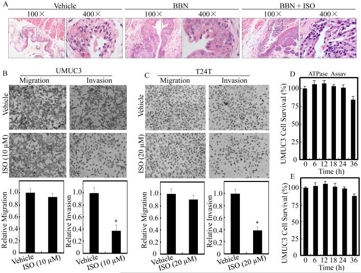
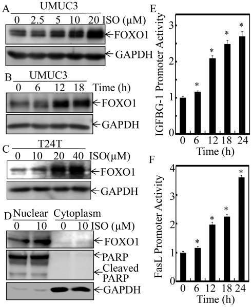
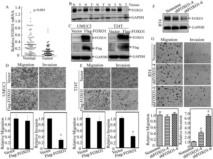
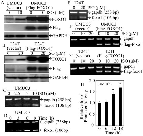
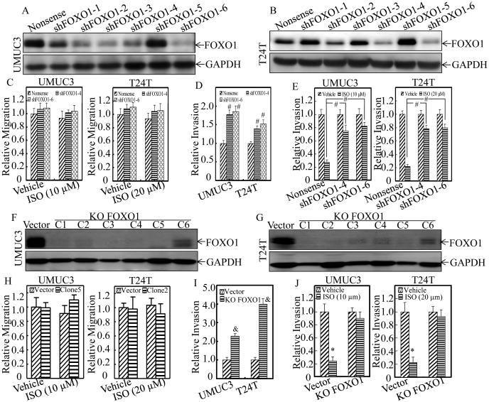
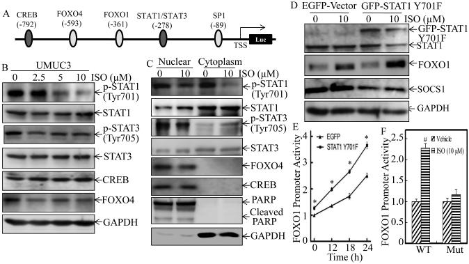
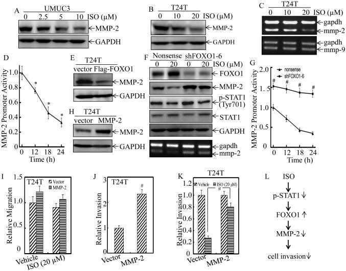
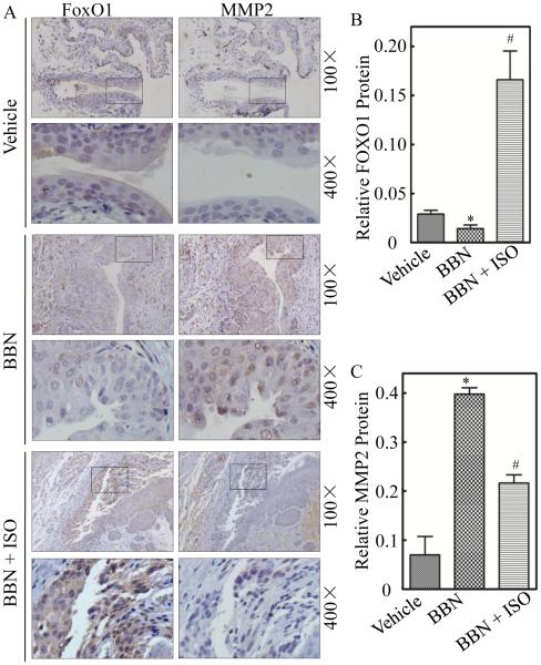
Although our most recent studies have identified Isorhapontigenin (ISO), a novel derivative of stilbene that isolated from a Chinese herb Gnetum cleistostachyum, for its inhibition of human bladder cancer growth, nothing is known whether ISO possesses an inhibitory effect on bladder cancer invasion. Thus, we addressed this important question in current study and discovered that ISO treatment could inhibit mouse-invasive bladder cancer development following bladder carcinogen N-butyl-N-(4-hydroxybutyl) nitrosamine (BBN) exposure in vivo We also found that ISO suppressed human bladder cancer cell invasion accompanied by upregulation of the forkhead box class O 1 (FOXO1) mRNA transcription in vitro Accordingly, FOXO1 was profoundly downregulated in human bladder cancer tissues and was negatively correlated with bladder cancer invasion. Forced expression of FOXO1 specifically suppressed high-grade human bladder cancer cell invasion, whereas knockdown of FOXO1 promoted noninvasive bladder cancer cells becoming invasive bladder cancer cells. Moreover, knockout of FOXO1 significantly increased bladder cancer cell invasion and abolished the ISO inhibition of invasion in human bladder cancer cells. Further studies showed that the inhibition of Signal transducer and activator of transcription 1 (STAT1) phosphorylation at Tyr701 was crucial for ISO upregulation of FOXO1 transcription. Furthermore, this study revealed that metalloproteinase-2 (MMP-2) was a FOXO1 downstream effector, which was also supported by data obtained from mouse model of ISO inhibition BBN-induced mouse-invasive bladder cancer formation. These findings not only provide a novel insight into the understanding of mechanism of bladder cancer's propensity to invasion, but also identify a new role and mechanisms underlying the natural compound ISO that specifically suppresses such bladder cancer invasion through targeting the STAT1-FOXO1-MMP-2 axis. Cancer Prev Res; 9(7); 567-80. ©2016 AACR.


Figures

Figure 1
Figure 1. ISO inhibited BC invasion in vivo and in vitro
(A) Mice were divided into vehicle-treated control group (n=12), BBN-treated group (n=12), and BBN combined with ISO-treated group (n=12). HE staining was performed and the representative images of each group were shown. Human UMUC3 (B) and T24T (C) BC cells were cultured in chamber or pre-coated matrigel chamber and treated with medium containing either vehicle or indicated concentration of ISO for 24 hours. The cells were then fixed and stained. The invasion and migration rates were quantified by counting the relative migrated (transwell) and invaded cells at least three random fields under a light microscope. (D) UMUC3 cells were treated with medium containing either vehicle or 10 µM ISO up to 36 hours for cell viability assessment by ATPase assay. (E) T24T cells were treated with medium containing either vehicle or 20 µM ISO for the indicated time. Cell viability was also evaluated by ATPase assay. Results are the means ± SD of triplicates. Symbol “*” indicates a significant difference between vehicle and ISO-treated group (P < 0.05).
Figure 2
Figure 2. ISO treatment up-regulated FOXO1 expression and enhanced FOXO1-dependent transcriptional activity in human BC cells
UMUC3 (A & B) and T24T cells (C) were treated with medium containing either vehicle or ISO as the indicated concentrations for 12 hours (A & C) or treated with medium containing either vehicle or 10 μM ISO for 6-18 hours as indicated (B). The whole cell lysates were used for Western blotting. GAPDH was used as protein loading control. (D) The cell nuclear and cytoplasm fractions of UMUC3 cells treated with 10 μM ISO for 12 hours were subjected to SDS-PAGE and immunoblotted with the indicated specific antibodies. The expressions of PARP and GAPDH were respectively used as markers for nuclear and cytoplasm fractions, respectively. (E) UMUC3 cells stably transfected with IGFBG-1 promoter-driven luciferase reporter was treated with 10 μM of ISO for the indicated times. Luciferase activity was determined by the Dual-Luciferase Reporter Assay System. (F) UMUC3 cells were stably transfected with FasL promoter-driven luciferase reporter construct, and was treated with10 μM of ISO for the indicated times to determine the promoter transcriptional activity. Results are the means ± SD of triplicates. Symbol “*” indicates a significant difference between vehicle and ISO-treated group (P < 0.05).
Figure 3
Figure 3. FOXO1 was down-regulated in human BC tissues and acted as a regulator of human BC cell invasion
Total RNA and protein lysates were prepared from human normal (N) and paired cancerous (T) tissues among 98 patients diagnosed with BCs, and subjected to Quantitative RT-PCR and Western blotting analyses for determining FOXO1 mRNA (A) and protein (B) expression profiles, respectively. Flag-FOXO1 expression construct was used to stably transfected into UMUC3 and T24T cells, respectively. The stable transfectants, UMUC3(Flag-FOXO1) and T24T(Flag-FOXO1) were identified by Western blotting (C), and then used for determination of their abilities of cell invasion and migration as compared with their vector control transfectants (D & E) as described in “Materials and Methods” section. (F) RT4 cells were stably transfected with Nonsense shRNA or two FOXO1 shRNA constructs (shFOXO1-4, shFOXO1-6), respectively, and the knockdown efficiency of FOXO1 protein was evaluated by Western Blot. (G) FOXO1 stably knockdown transfectants, RT4(shFOXO1-4) and RT4(shFOXO1-6), as well as Nonsense transfectant RT4(Nonsense) were then used for determination of their migration and invasion abilities. Results are the means ± SD of triplicates. Symbol “*” indicates a significant difference between vector control and FOXO1 overexpression group (P < 0.05). Symbol “#” indicates a significant difference between Nonsense transfectant and shRNA transfectants (P < 0.05).
Figure 4
Figure 4. ISO up-regulated FOXO1 expression at mRNA transcriptional level in human BC cells
UMUC3(Vector) and UMUC3(Flag-FOXO1) (A) or T24T(Vector) and T24T(Flag-FOXO1) (B) cells were treated with medium containing either vehicle or ISO as the indicated concentrations for 12 hours. The cell extracts were subjected to Western Blotting for determination of ISO on protein expression of FOXO1 and Flag-FOXO1 as indicated. UMUC3 (C & D) and T24T (E) cells were treated with ISO for 6 hours at indicated concentrations (C & E) or 10 μM ISO for the indicated time (D), and foxo1 mRNA expression was determined by RT-PCR. The gapdh mRNA level was used as loading control. UMUC3(Vector) and UMUC3(Flag-FOXO1) (F) or T24T (Vector) and T24T (Flag-FOXO1) (G) cells were treated with medium containing either vehicle or ISO as the indicated concentrations for 6 hours. The Flag-foxo1 mRNA levels were evaluated by RT-PCT as indicated. (H) UMUC3 cells stably transfected with foxo1 promoter-driven luciferase reporter was treated with 10 μM ISO for the indicated times. Luciferase activity was evaluated by the Dual-Luciferase Reporter Assay System. Results are the means ± SD of triplicates. Symbol “*” indicates a significant difference between vehicle and ISO-treated group (P < 0.05).
Figure 5
Figure 5. Knockdown and knockout of FOXO1 reversed ISO inhibition of BC cell invasion
(A-E) UMUC3 (A) and T24T (B) cells were stably transfected with Nonsense shRNA or a set of six various FOXO1 shRNA (shFOXO1) constructs, respectively. The knockdown efficiency of FOXO1 protein was assessed by western blotting. FOXO1 stably knockdown transfectants, including UMUC3(shFOXO1-4), UMUC3(shFOXO1-6), T24T(shFOXO1-4) and T24T(shFOXO1-6), as well as Nonsense transfectants were then used for determination of their cell migration (C) and invasion (D & E) in presence of either vehicle or the indicated concentration of ISO treatment for 24 hours. (F-J) CRISPR/Cas9 systems were then applied to knockout FOXO1 gene, and the single clone stable FOXO1 knockout transfectant, UMUC3(KO FOXO1, C1-C6) and T24T(KO FOXO1, C1-C6), were identified by Western Blot (F & G). FOXO1 stably knockout transfectants, including UMUC3(KO FOXO1, clone5) and T24T(KO FOXO1, clone2), as well as Vector control transfectants were used for determination of their cell migration (H) and invasion (I & J) in presence of either vehicle or the indicated concentration of ISO treatment for 24 hours. Results are the means ± SD of triplicates. Symbol “#” indicates a significant difference between Nonsense transfectant and shRNA transfectants (P < 0.05). Symbol “&” indicates a significant difference between scramble vector control and FOXO1 knockout transfectants (P<0.05). Symbol “*” indicates a significant difference between vehicle and ISO-treated group (P < 0.05).
Figure 6
Figure 6. ISO promoted FOXO1 transcription by inhibition of STAT1 phosphorylation at Tyr701
(A) Schematic representation of transcription factor binding sites of human FOXO1 gene promoter. (B) UMUC3 cells were treated with either vehicle or ISO as the indicated concentrations for 12 hours. Expression of the related transcription factors in the whole cell lysates was determined by Western blotting, and GAPDH was used as protein loading control. (C) The UMUC3 cells were treated with either vehicle or 10 μM of ISO for 12 hours. The cell extracts were used to isolate cell nuclear and cytoplasm fractions and then subjected to Western Blotting with the specific antibodies as indicated. PARP and GAPDH were used as markers for nuclear and cytoplasm fractions, respectively. (D) UMUC3 cells were stably transfected with dominant negative STAT1, GFP-STAT1 Y701F, and the stable transfectants were treated with either vehicle or 10 μM ISO for 12 hours. The expression of FOXO1 and SOCS1 was determined by Western blotting. GAPDH was used as protein loading control. (E) FOXO-1 promoter-driven luciferase reporter was tranfected into UMUC3(EGFP) and UMUC3(STAT1 Y701F) cells, and the stable transfectants were then treated with 10 μM of ISO for the indicated times. Luciferase activity was evaluated by the Dual-Luciferase Reporter Assay System. (F) UMUC3 cells were stably transfected with FOXO-1 promoter-driven luciferase or the STAT1 binding site mutant, and the stable transfectants were treated with 10 μM of ISO for 18 hours. Dual-Luciferase Reporter Assay System was performed to determine the Luciferase activity. Results are the means ± SD of triplicates. Symbol “*” indicates a significant difference between UMUC3(EGFP-vector) and UMUC3(GFP-STAT1 Y701F) in the same time point upon ISO treatment (P <0.05). Symbol “#”indicates a significant difference between vehicle control and ISO–treated group (P< 0.05).
Figure 7
Figure 7. ISO inhibited BC cell invasion through down-regulation of MMP-2 in a FOXO1-denpendent manner
UMUC3 (A) and T24T (B) cells were treated with either vehicle or ISO as the indicated concentrations for 18 hours. The protein level of MMP-2 was determined by Western blotting, and GAPDH was used as protein loading control. (C) T24T cells were treated with either vehicle or the indicated concentrations of ISO for 18 hours. The mmp-2 and mmp-9 mRNA levels were evaluated by RT-PCR, and gapdh mRNA was used as the internal loading control. (D) T24T cells were stably transfected with MMP-2 promoter-driven luciferase reporter and the stable transfectants were treated with 20 μM of ISO for the indicated times. Dual-Luciferase Reporter Assay System was used to detect the Luciferase activity. Results are the means ± SD of triplicates. Symbol “*” indicates a significant difference between vehicle control and ISO-treated groups (P < 0.05). (E) The expression of FOXO1 in T24T(Vector) and T24T(Flag-FOXO1) stable transfectants were detected by Western blotting. (F) T24T(shFOXO1-6) and T24T(Nonsense) cells were treated with vehicle or 20 μM of ISO for 18 hours. Cell lysates were subjected to Western blotting with the specific antibodies indicated. (G) T24T(shFOXO1-6) and T24T(Nonsense) cells were tranfected with MMP-2 promoter-driven luciferase reporter, and the stable transfectants were then treated with 20 μM of ISO for the indicated times, and cells were then subjected to determine luciferase activity using Dual-Luciferase Reporter Assay. Results are the means ± SD of triplicates. Symbol “#” indicates a significant difference between T24T(shFOXO1-6) and T24T(Nonsense) cells in the same time point upon ISO treatment (P < 0.05). (H) MMP-2 was stably transfected into T24T cells and the stable transfectant was identified by verification of ectopic expression of MMP-2 protein by Western blotting. The MMP-2 stable transfectant and its vector control transfectant were subjected to cell migration (I) and invasion (J & K) assay in presence of either vehicle or 20 μM of ISO for 24 hours. Results are the means ± SD of triplicates. Symbol “#” indicates a significant difference between Vector transfectant and MMP-2 stable transfectant (P<0.05). (L) The proposed schematic for the cascade underlying ISO inhibition of human BC cell invasion through down-regulation of MMP-2 in FOXO1-dependent manner.
Figure 8
Figure 8. ISO reversed the BBN-induced down-regulation of FOXO1 and up-regulation of MMP-2 in mice
Mice were divided into vehicle-treated control group (n=12), BBN-treated group (n=12), and BBN combined with ISO-treated group (n=12). (A) IHC-P staining by antibodies specific against FOXO1 and MMP-2 were performed. FOXO1 (B) and MMP-2 (C) protein expression levels were analyzed by calculating the integrated optical density per stained area (IOD/area) using Image-Pro Plus version 6.0. Results are the means ± SD of 12 mice in each group. Symbol “*” indicates a significant difference between vehicle control group and BBN-treated group (P < 0.05). Symbol “#” indicates a significant difference between BBN-treated group and BBN combined with ISO-treated group (P < 0.05).

Similar articles

Cited by

References

    1. Siegel R, Ma J, Zou Z, Jemal A. Cancer statistics, 2014. CA Cancer J Clin. 2014;64:9–29. - PubMed
    1. Kaufman DS, Shipley WU, Feldman AS. Bladder cancer. Lancet. 2009;374:239–49. - PubMed
    1. Huang KS, Wang YH, Li RL, Lin M. Stilbene dimers from the lianas of Gnetum hainanense. Phytochemistry. 2000;54:875–81. - PubMed
    1. Fang Y, Cao Z, Hou Q, Ma C, Yao C, Li J, et al. Cyclin d1 downregulation contributes to anticancer effect of isorhapontigenin on human bladder cancer cells. Mol Cancer Ther. 2013;12:1492–503. - PMC - PubMed
    1. Fang Y, Yu Y, Hou Q, Zheng X, Zhang M, Zhang D, et al. The Chinese herb isolate isorhapontigenin induces apoptosis in human cancer cells by down-regulating overexpression of antiapoptotic protein XIAP. J Biol Chem. 2012;287:35234–43. - PMC - PubMed

MeSH terms