

The first see-through frog created by breeding: description, inheritance patterns, and dermal chromatophore structure
- PMID: 27080918
- PMCID: PMC4832234
- DOI: 10.1038/srep24431
The first see-through frog created by breeding: description, inheritance patterns, and dermal chromatophore structure
Abstract
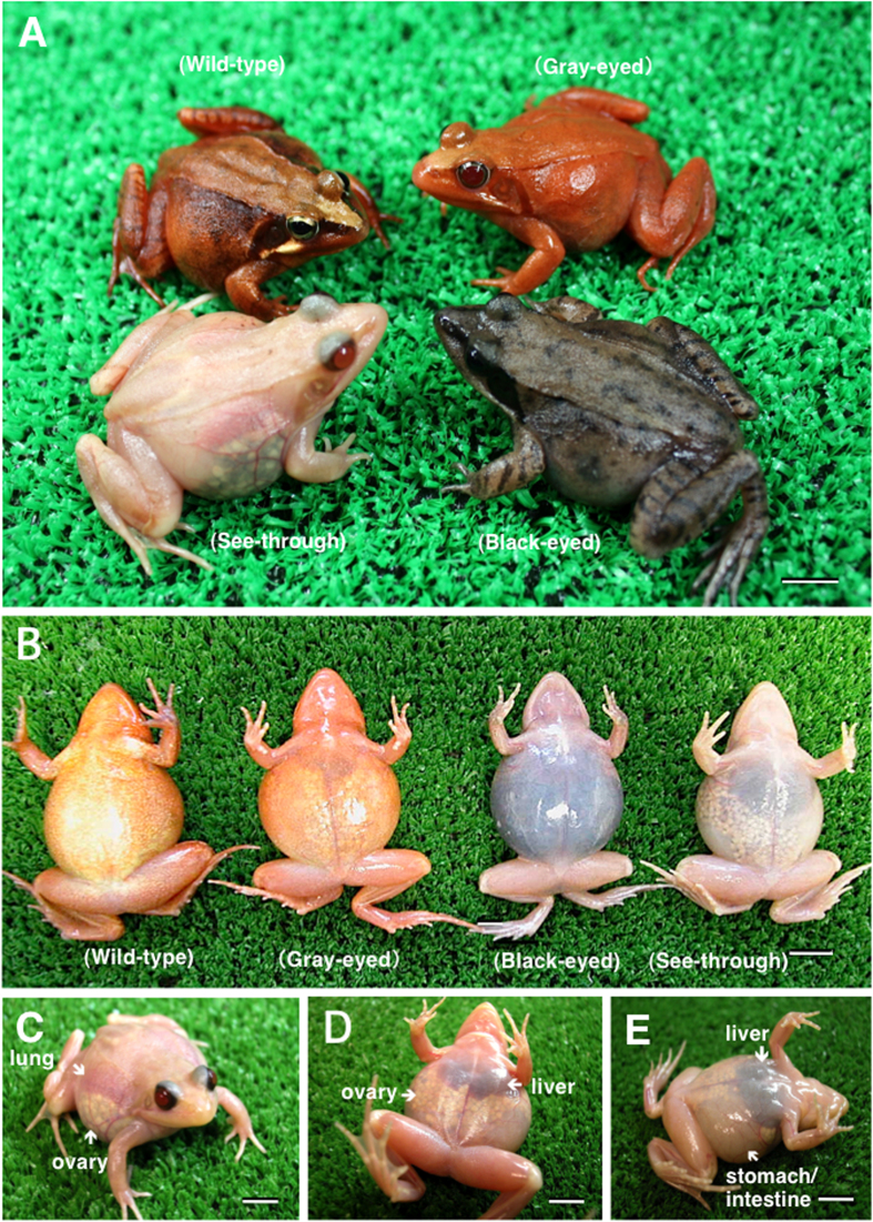
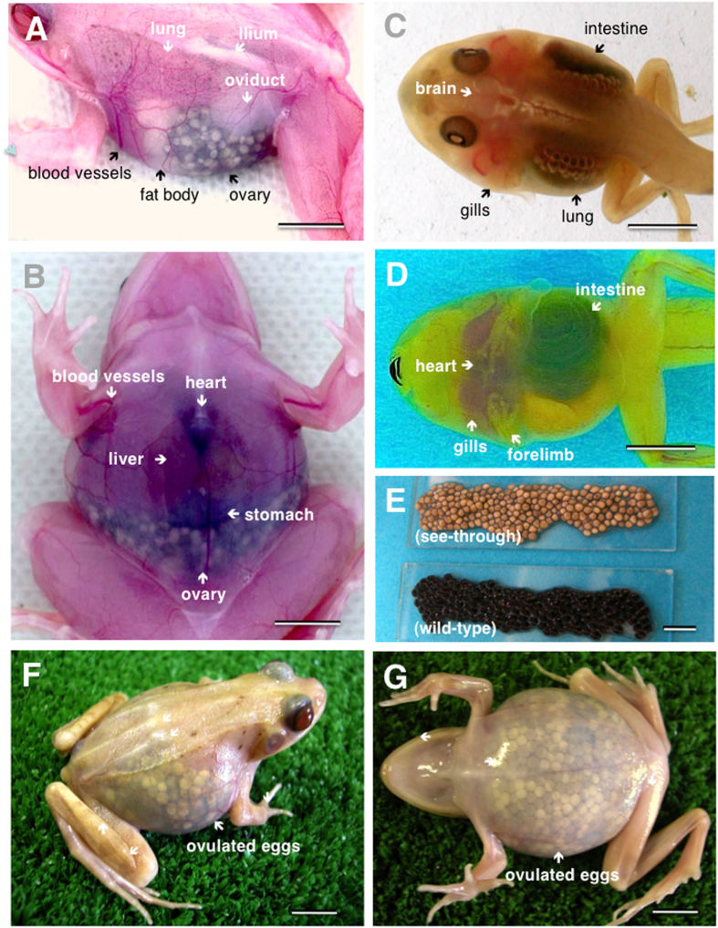
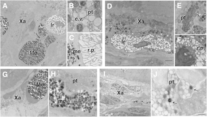
We have succeeded in creating see-through frogs from natural color mutants of the Japanese brown frog Rana japonica, which usually possesses an ochre or brown back; this coloration enables the organs, blood vessels, and eggs to be observed through the skin without performing dissection. We crossed two kinds of recessive color mutant (black-eyed and gray-eyed) frogs through artificial insemination, and F2 offspring produced frogs whose skin is translucent throughout the life cycle. Three kinds of dermal chromatophores--xanthophores, iridophores, and melanophores--are observed in a layered arrangement in the skin of wild-type frogs, but few chromatophores were present in the skin of the see-through frogs. The translucent skin enables observation of organ growth and cancer formation and progression in the animal, which can be monitored over its entire life without the need for dissection. See-through frogs thus provide a useful animal model for environmental, medical, and biological research.
Figures
Similar articles
-
Genetic and experimental studies on a pigment mutation, Pale (Pa), in the frog, Bombina orientalis.J Embryol Exp Morphol. 1980 Apr;56:125-37. J Embryol Exp Morphol. 1980. PMID: 7400738
-
The dermal chromatophore unit.J Cell Biol. 1968 Jul;38(1):67-79. doi: 10.1083/jcb.38.1.67. J Cell Biol. 1968. PMID: 5691979 Free PMC article.
-
Ultrastructure of the dermal chromatophores in a lizard (Scincidae: Plestiodon latiscutatus) with conspicuous body and tail coloration.Zoolog Sci. 2006 Sep;23(9):793-9. doi: 10.2108/zsj.23.793. Zoolog Sci. 2006. PMID: 17043401
-
Pigment pattern formation in zebrafish: a model for developmental genetics and the evolution of form.Microsc Res Tech. 2002 Sep 15;58(6):442-55. doi: 10.1002/jemt.10162. Microsc Res Tech. 2002. PMID: 12242701 Review.
-
The physiology of flatfish chromatophores.Microsc Res Tech. 2002 Sep 15;58(6):481-7. doi: 10.1002/jemt.10166. Microsc Res Tech. 2002. PMID: 12242705 Review.
Cited by
-
Glassfrogs conceal blood in their liver to maintain transparency.Science. 2022 Dec 23;378(6626):1315-1320. doi: 10.1126/science.abl6620. Epub 2022 Dec 22. Science. 2022. PMID: 36548427 Free PMC article.
-
Revealing mitf functions and visualizing allografted tumor metastasis in colorless and immunodeficient Xenopus tropicalis.Commun Biol. 2024 Mar 5;7(1):275. doi: 10.1038/s42003-024-05967-3. Commun Biol. 2024. PMID: 38443437 Free PMC article.
-
It's not easy being green: Comparing typical skin colouration among amphibians with colour abnormalities associated with chromatophore deficits.Ecol Evol. 2024 May 21;14(5):e11438. doi: 10.1002/ece3.11438. eCollection 2024 May. Ecol Evol. 2024. PMID: 38779532 Free PMC article.
References
-
- Akiyama S.-I. & Tamaru Y. Development of transgenic technology using transparent goldfish. Abst. Ann. Meet. Soc. Biosci. Bioengineer. Japan 2010, 19 (2010).
-
- Twomey E., Delia J. & Castroviejo-Fisher S. A review of Northern Peruvian glassfrogs (Centrolenidae), with the description of four new remarkable species. Zootaxa 3851, 1–87 (2014). - PubMed
-
- Kubicki B., Salazar S. & Puschendorf R. A new species of glassfrog, genus Hyalinobatrachium (Anura: Centrolenidae), from the Caribbean foothills of Costa Rica. Zootaxa 3920, 69–84 (2015). - PubMed
Publication types
MeSH terms
LinkOut - more resources
Full Text Sources
Other Literature Sources
Miscellaneous
