. 2016 Apr 14:17:67.
doi: 10.1186/s13059-016-0932-1.

Single-cell analysis of long non-coding RNAs in the developing human neocortex

Affiliations

Single-cell analysis of long non-coding RNAs in the developing human neocortex

Siyuan John Liu et al. Genome Biol. .

Abstract

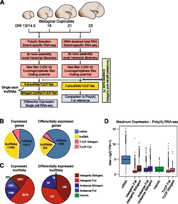
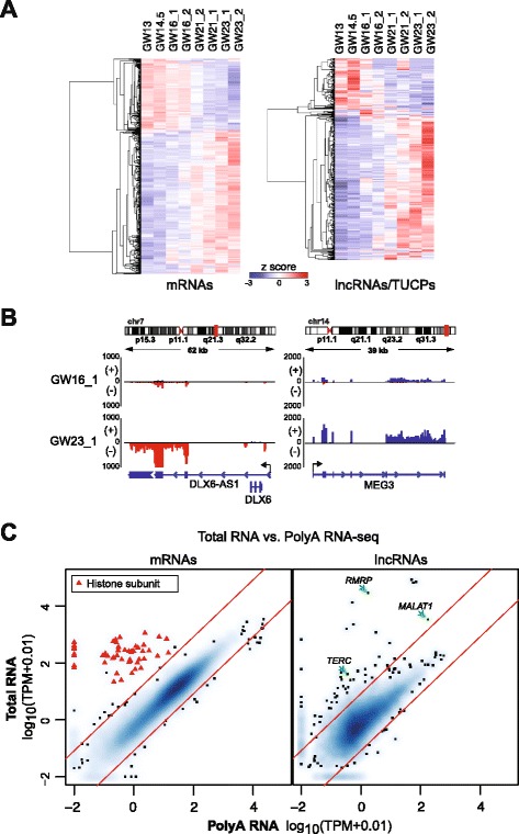
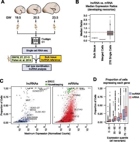
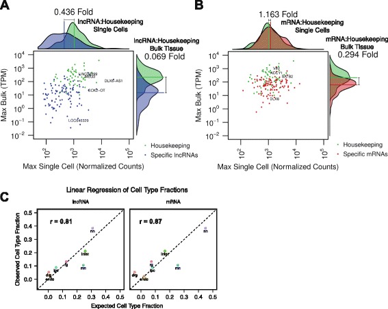
Background: Long non-coding RNAs (lncRNAs) comprise a diverse class of transcripts that can regulate molecular and cellular processes in brain development and disease. LncRNAs exhibit cell type- and tissue-specific expression, but little is known about the expression and function of lncRNAs in the developing human brain. Furthermore, it has been unclear whether lncRNAs are highly expressed in subsets of cells within tissues, despite appearing lowly expressed in bulk populations.

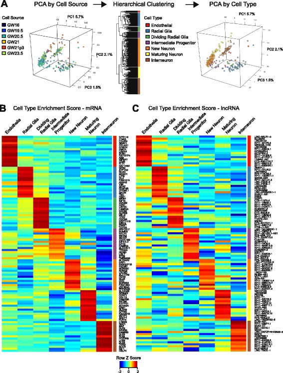
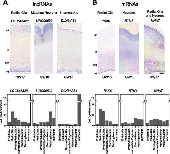
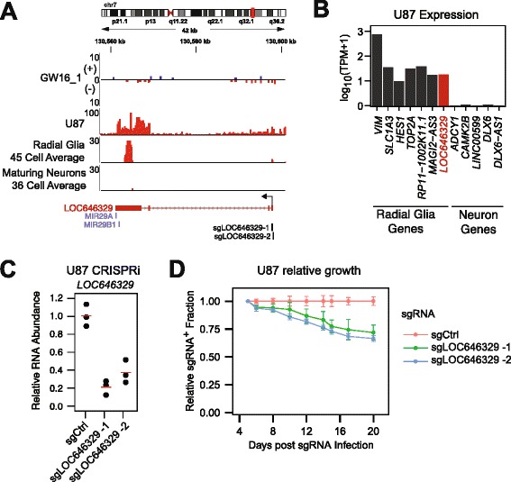
Results: We use strand-specific RNA-seq to deeply profile lncRNAs from polyadenylated and total RNA obtained from human neocortex at different stages of development, and we apply this reference to analyze the transcriptomes of single cells. While lncRNAs are generally detected at low levels in bulk tissues, single-cell transcriptomics of hundreds of neocortex cells reveal that many lncRNAs are abundantly expressed in individual cells and are cell type-specific. Notably, LOC646329 is a lncRNA enriched in single radial glia cells but is detected at low abundance in tissues. CRISPRi knockdown of LOC646329 indicates that this lncRNA regulates cell proliferation.

Conclusion: The discrete and abundant expression of lncRNAs among individual cells has important implications for both their biological function and utility for distinguishing neural cell types.

Keywords: CRISPRi; Developing brain; Single-cell RNA-seq; lncRNA.


Figures

Fig. 1
Fig. 1
Catalogue of lncRNAs in human neocortex development. a Schematic of neocortex tissue dissection, poly(A) and total RNA-seq library prep, and computational pipeline for lncRNA annotation and quantification. b Numbers of expressed (left) and differentially expressed (right; DESeq2, FDR <0.01) mRNAs, lncRNAs, and TUCPs during neocortex development in bulk tissues. Stringent references omit novel single exon transcripts. c Breakdown of expressed (left) and differentially expressed (right) lncRNAs based on genomic orientation relative to mRNAs. d Maximum expression levels of transcripts described in the Full and Stringent references derived from Poly(A) selection RNA-seq, across all samples. TPM, Transcripts per Million
Fig. 2
Fig. 2
Differential expression of mRNAs and lncRNAs/TUCPs during neocortex development. a Heatmaps of differentially expressed mRNAs (left) and lncRNAs/TUCPs (right) throughout eight samples of bulk neocortex tissues. b Strand-specific RNA-seq alignments at the DLX6-AS1 and MEG3 loci in GW16 and GW23 replicate one sample. Scale, number of aligned reads. c Comparison of mRNA (left) and lncRNA (right) expression levels between poly(A) RNA-seq and total RNA-seq in GW16 sample 2. Red diagonals represent 10-fold enrichment in either total (upper) or polyA (lower) fractions. Red triangles, histone subunits enriched >10-fold in total RNA. TPM, Transcripts per Million
Fig. 3
Fig. 3
Single-cell transcriptomics of lncRNA expression. a Schematic of single-cell microfluidic capture and integration of transcriptome reference generated from bulk tissue RNA-seq to conduct cell-type identification and lncRNA analysis. Previously captured cells from Pollen et al. [32] were also included. b Distributions of median lncRNA expression to median mRNA expression ratios (lncRNA:mRNA) in bulk tissues, in silico merged single cells, and single cells from the developing neocortex. c Proportion of neocortex cells that expressed each lncRNA (blue) and mRNA (red), separated by maximum expression in single cells. d Same as in (c) but grouped by maximum expression quantile of the set of all transcripts (lncRNA and mRNA combined). Green squares, housekeeping genes; black triangles, ERCC Spike-In Controls
Fig. 4
Fig. 4
Cell type-specific expression of lncRNAs. a Identifying cell types using unsupervised clustering. Left – Principal component analysis (PCA) of single cells colored by developmental stage of source tissues. Middle – Complete linkage hierarchical clustering of single cells using genes exhibiting variance greater than expected than from technical noise. Right – PCA of single cells colored by cell types inferred from protein coding genes specific to each cluster. Axes labels indicate percent variation explained by each PC. b Heatmaps of cell type enrichment scores for the 15 most specific mRNAs and (c) lncRNAs in each cluster. GW21p3, primary cells derived from GW21 brain that were cultured in differentiation media for 3 days
Fig. 5
Fig. 5
In situ hybridization of cell type-specific lncRNAs and mRNAs in developing neocortex. a In situ hybridizations and corresponding cell type enrichment values for radial glia-specific lncRNA LOC646329 (left), maturing neuron-specific lncRNA LINC00599 (middle), and interneuron-specific lncRNA DLX6-AS1 (right). b In situ hybridizations and corresponding cell type enrichment values for radial glia-specific mRNA PAX6 (left), neuron mRNA marker RTN1 (middle), and progenitor and differentiated cell-expressed mRNA NNAT (right). Scale bars, 250 μm. CP, cortical plate. IZ, intermediate zone. SVZ, subventricular zone. VZ, ventricular zone
Fig. 6
Fig. 6
Cell type-specific lncRNAs appear to be lowly expressed in bulk tissues. a Comparison of single-cell and bulk tissue maximum expression levels of 105 cell type-specific lncRNAs and (b) 105 cell type-specific mRNAs. Green, housekeeping genes; blue, cell type-specific lncRNAs; red, cell type-specific mRNAs. Projected density plots summarize expression levels of scatterplots along the single-cell (horizontal) and bulk tissue (vertical) axes. Fold changes noted alongside the projected density plots represent the ratio of the median expression of cell type-specific lncRNAs or mRNAs to the median expression of housekeeping genes in single cell or whole tissue RNA-seq. c Comparison of expected cell type fractions as predicted by linear regression (x axis) and observed cell type fractions (y axis). TPM, Transcripts per million; Endo, endothelial; rg, radial glia; drg, dividing radial glia; ipc, intermediate progenitor cell; nn, newborn neurons; mn, maturing neurons; inter, interneurons
Fig. 7
Fig. 7
CRISPRi knockdown of radial glia-enriched lncRNA LOC646329 inhibits proliferation. a RNA-seq alignments at the LOC646329 locus. GW16(+)/(–) replicate one and U87 alignments are number of reads. Radial glia and maturing neurons are merged alignments normalized by number of cells within each cell cluster. sgRNAs targeting the TSS of LOC646329 are indicated. b Expression of radial glia and neuron mRNAs and lncRNAs in U87 glioblastoma cells. c qPCR of LOC646329 following 4 days of CRISPRi knockdown using two sgRNAs targeting the TSS of LOC646329 relative to non-targeting control sgRNA. Biological triplicates (black circles) show 79.0 % repression with sgLOC646329-1 (p = 0.0014; Welch’s t-test) and 62.6 % repression in sgLOC646329-2 (p = 0.0035; Welch’s t-test). Red lines, mean. d Relative growth assays of U87 cells following sgRNA infection. sgRNA+ fraction was calculated relative to 5 days post sgRNA infection and normalized to the sgCtrl+ fraction at each time point. Biological triplicates show 28.1 % depletion at 20 days with sgLOC646329-1 (p = 0.0073; Welch’s t-test) and 33.5 % depletion with sgLOC646329-2 (p = 0.00048; Welch’s t-test). Error bars, standard deviation of triplicate cultures

Comment in

References

    1. Sauvageau M, Goff LA, Lodato S, Bonev B, Groff AF, Gerhardinger C, et al. Multiple knockout mouse models reveal lincRNAs are required for life and brain development. eLife. 2013;2:e01749. doi: 10.7554/eLife.01749. - DOI - PMC - PubMed
    1. Goff LA, Groff AF, Sauvageau M, Trayes-Gibson Z, Sanchez-Gomez DB, Morse M, et al. Spatiotemporal expression and transcriptional perturbations by long noncoding RNAs in the mouse brain. Proc Natl Acad Sci. 2015;112:6855–62. doi: 10.1073/pnas.1411263112. - DOI - PMC - PubMed
    1. Bond AM, VanGompel MJW, Sametsky EA, Clark MF, Savage JC, Disterhoft JF, et al. Balanced gene regulation by an embryonic brain ncRNA is critical for adult hippocampal GABA circuitry. Nat Neurosci. 2009;12:1020–7. doi: 10.1038/nn.2371. - DOI - PMC - PubMed
    1. Cabili MN, Trapnell C, Goff L, Koziol M, Tazon-Vega B, Regev A, et al. Integrative annotation of human large intergenic noncoding RNAs reveals global properties and specific subclasses. Genes Dev. 2011;25:1915–27. doi: 10.1101/gad.17446611. - DOI - PMC - PubMed
    1. Ramos AD, Diaz A, Nellore A, Delgado RN, Park K-Y, Gonzales-Roybal G, et al. Integration of genome-wide approaches identifies lncRNAs of adult neural stem cells and their progeny in vivo. Cell Stem Cell. 2013;12:616–28. doi: 10.1016/j.stem.2013.03.003. - DOI - PMC - PubMed

Publication types

Substances