. 2016 Jun;15(6):2125-40.
doi: 10.1074/mcp.M115.056440. Epub 2016 Apr 14.

Comprehensively Characterizing the Thioredoxin Interactome In Vivo Highlights the Central Role Played by This Ubiquitous Oxidoreductase in Redox Control

Affiliations

Comprehensively Characterizing the Thioredoxin Interactome In Vivo Highlights the Central Role Played by This Ubiquitous Oxidoreductase in Redox Control

Isabelle S Arts et al. Mol Cell Proteomics. 2016 Jun.

Abstract

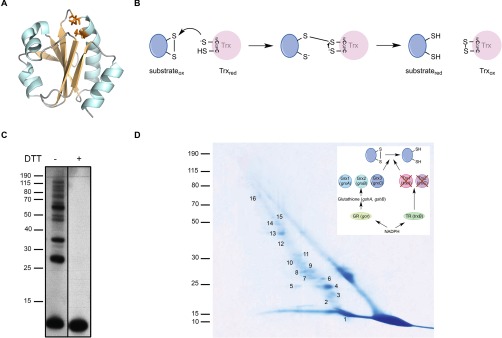
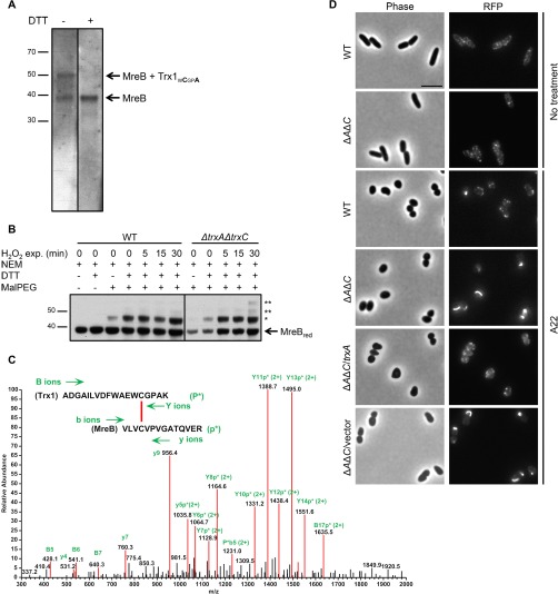
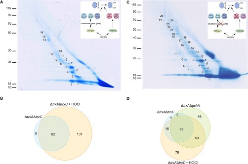
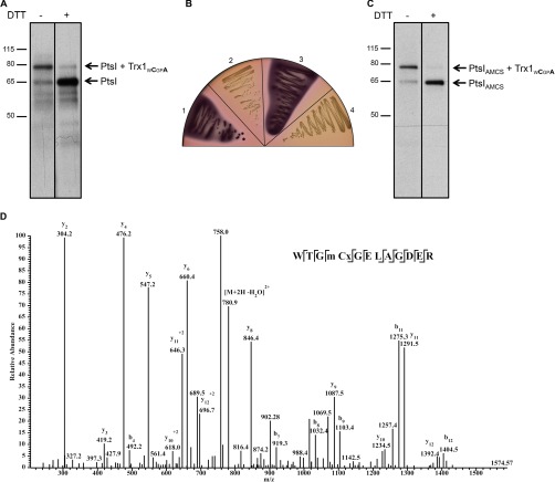
Thioredoxin (Trx) is a ubiquitous oxidoreductase maintaining protein-bound cysteine residues in the reduced thiol state. Here, we combined a well-established method to trap Trx substrates with the power of bacterial genetics to comprehensively characterize the in vivo Trx redox interactome in the model bacterium Escherichia coli Using strains engineered to optimize trapping, we report the identification of a total 268 Trx substrates, including 201 that had never been reported to depend on Trx for reduction. The newly identified Trx substrates are involved in a variety of cellular processes, ranging from energy metabolism to amino acid synthesis and transcription. The interaction between Trx and two of its newly identified substrates, a protein required for the import of most carbohydrates, PtsI, and the bacterial actin homolog MreB was studied in detail. We provide direct evidence that PtsI and MreB contain cysteine residues that are susceptible to oxidation and that participate in the formation of an intermolecular disulfide with Trx. By considerably expanding the number of Trx targets, our work highlights the role played by this major oxidoreductase in a variety of cellular processes. Moreover, as the dependence on Trx for reduction is often conserved across species, it also provides insightful information on the interactome of Trx in organisms other than E. coli.


Figures

Fig. 1.
Fig. 1.
Trapping of Trx1 substrates in a ΔtrxAΔtrxC E. coli strain in vivo. A, Structure of reduced E. coli thioredoxin (PDB code: 1XOB). All thioredoxins share a canonical WCGPC catalytic motif located on a highly conserved fold, which consists of five β-strands (light orange) surrounded by four α-helices (cyan). The cysteines of the catalytic motif are represented in orange. The figure was generated using MacPyMol (Delano Scientific LLC 2006). B, Reaction mechanism of disulfide reduction by thioredoxin. The reaction takes off with a nucleophilic attack of the N-terminal cysteine of the conserved WCGPC motif targeting the disulfide. As a result, an intermediate mixed disulfide complex is formed between Trx and the substrate protein, which in turn is reduced by a nucleophilic attack of the C-terminal cysteine of the WCGPC motif. C, To trap proteins linked to Trx1, Trx1WCGPA was expressed in a ΔtrxAΔtrxC mutant. The proteins were TCA-precipitated and analyzed by Western blotting using an anti-His antibody. Induction of Trx1WCGPA led to the accumulation of several high-molecular weight complexes, which disappeared following addition of DTT. The molecular mass markers (in kDa) are indicated on the left. D, The mixed-disulfide complexes were purified by affinity chromatography using Ni-NTA agarose. After concentration, the proteins trapped by Trx1WCGPA were separated by SDS-PAGE (nonreducing dimension). The complexes were then separated in a second, reducing dimension. Proteins were identified by mass spectrometry. The spots analyzed by mass spectrometry were numbered as indicated (see Table I). The molecular mass markers (in kDa) are indicated on the left. The inset shows the reducing pathway that was genetically modified. Here, by deleting the trxA and trxC genes, it is the Trx pathway that was impaired. Trx, thioredoxin; TR, thioredoxin reductase; Grx, glutaredoxin; gshA encodes the gamma-glutamylcysteine synthetase; gshB encodes the glutathione synthetase; GR, glutaredoxin reductase.
Fig. 2.
Fig. 2.
The actin homolog MreB is a Trx1 substrate. A, After expression of Trx1WCGPA in a ΔtrxAΔtrxC mutant and purification and concentration of the complexes, the elution fraction was analyzed by Western blotting using an anti-MreB antibody. The bands corresponding to MreB alone or in complex with Trx1WCGPA are indicated. Upon DTT addition, the band corresponding to the complex disappeared whereas the MreB band increased, indicating that Trx1WCGPA is released from MreB under reducing conditions. The molecular mass markers (in kDa) are indicated on the left. The fact that MreB was observed in the nonreduced sample indicates that a portion of this protein co-purified with Trx1WCGPA on the Ni-NTA agarose column. B, To determine if MreB can become oxidized in vivo, a reverse thiol trapping experiment was performed in a wild-type (WT) strain and a ΔtrxAΔtrxC mutant strain. All reduced cysteine residues were first covalently modified with NEM. Oxidized cysteines are then reduced with DTT and subsequently alkylated with MalPEG. MalPEG modifies free thiols leading to a major shift on SDS-PAGE. Samples were analyzed by Western blotting using an anti-MreB antibody. The band corresponding to reduced MreB (MreBred) is indicated. Upon MalPEG addition, a slow-migrating band (*) appears when samples from both the wild type and the ΔtrxAΔtrxC mutant are treated, indicating that MreB can be oxidized in vivo. Exposure (exp.) of cells to 1 mm H2O2 led to the appearance of two additional higher migrating bands (**) in the ΔtrxAΔtrxC mutant but not in the wild type, which indicates that MreB is further oxidized when the Trx system is impaired. The molecular mass markers (in kDa) are indicated on the left. Both parts of the figure come from a single membrane. C, Trx1WCGPA forms a mixed disulfide complex with MreB. Mass spectrometry analysis of a triply charged parent ion of [M+3H]3+ = 1139.3 Da shows fragmentation characteristics of a disulfide linkage between Cys33 of Trx and Cys113 of MreB, as determined by the DBond software (46). P*, one strand of a dipeptide; p*, the other strand of a dipeptide; capital letters, fragment ions from peptide P*; lowercase letters, fragment ions from peptide p*. D, Wild-type (WT) and ΔtrxAΔtrxCAΔC) cells expressing an MreB-RFP-MreB sandwich fluorescent fusion were observed using phase contrast (left) and fluorescence microscopy (right). MreB localized similarly in the ΔtrxAΔtrxC mutant and in the wild type when cells were grown under normal conditions. Upon A22 treatment, an abnormal MreB localization pattern was observed in the ΔtrxAΔtrxC double mutant but not in the wild type. Expression of Trx1 (but not of the empty plasmid) in trans restored an MreB normal localization pattern. Bar, 5 μm.
Fig. 3.
Fig. 3.
Trapping of Trx1 substrates in a ΔtrxAΔtrxC E. coli strain in the presence of HOCl and in a in a ΔtrxAΔgshA E. coli strain in vivo. A, To identify Trx1 substrates upon oxidative stress, ΔtrxAΔtrxC cells expressing Trx1WCGPA were exposed to HOCl for 10 min. The mixed-disulfide complexes were then purified by affinity chromatography using Ni-NTA agarose. After concentration, the proteins trapped by Trx1WCGPA were separated by SDS-PAGE (nonreducing dimension). The complexes were then separated in a second, reducing dimension. Proteins were identified by mass spectrometry. The spots analyzed by mass spectrometry were numbered as indicated (see supplemental Table S9). The molecular mass markers (in kDa) are indicated on the left. The inset shows the reducing pathway that was genetically modified. Here, by deleting the trxA and trxC genes, it is the Trx pathway that was impaired. Trx, thioredoxin; TR, thioredoxin reductase; Grx, glutaredoxin; gshA encodes the gamma-glutamylcysteine synthetase; gshB encodes the glutathione synthetase; GR, glutaredoxin reductase. B, Venn diagram representing the numbers of proteins identified in the ΔtrxAΔtrxC strain with or without HOCl stress (from Tables I and supplemental Table S9). C, To identify the Trx1 substrates that may have been missed because of the activity of the Grx pathway, Trx1WCGPA was expressed in a ΔtrxAΔgshA mutant. The mixed-disulfide complexes were then purified, concentrated and separated as described in A. Proteins were identified by mass spectrometry. The spots analyzed by mass spectrometry were numbered as indicated (see supplemental Table S10). The molecular mass markers (in kDa) are indicated on the left. The inset shows the reducing pathways that were genetically modified. Here, by deleting the trxA and gshA genes, both the Trx and the Grx pathways were impaired. Legend is the same as in A. D, Venn diagram representing the numbers of proteins identified in the ΔtrxAΔtrxC (with HOCl stress or not) and the ΔtrxAΔgshA strains (from Tables I, S9 and S10).
Fig. 4.
Fig. 4.
Identification of PtsI as a novel redox-regulated enzyme. A, Trx1WCGPA was expressed in a ΔtrxAΔgshA mutant, purified and concentrated. The elution fraction was then analyzed by Western blotting using an anti-PtsI antibody. The bands corresponding to PtsI alone or in complex with Trx1WCGPA are indicated. Upon DTT addition, the band corresponding to the complex between PtsI and Trx1 disappeared whereas the PtsI band increased, indicating that Trx1WCGPA is released from PtsI under reducing conditions. The molecular mass markers (in kDa) are indicated on the left. The fact that PtsI was observed in the nonreduced sample indicates that a portion of this protein co-purified with Trx1WCGPA on the Ni-NTA agarose column. B, Strains expressing different versions of PtsI were streaked on MacConkey plates (containing 0.5% glucose) and glucose fermentation was analyzed. Red colonies indicate glucose fermentation whereas white colonies show no fermentation. Strains are as follows: 1. Wild type, 2. ΔptsI, 3. ptsI::ptsIAMCS, 4. ptsI::ptsIC502S. Cells with a nonfunctional PtsI cannot ferment glucose because of an impaired glucose uptake. C, The complex formed between Trx1WCGPA and PtsIAMCS was analyzed as in A, in a ΔtrxAΔgshA mutant expressing PtsIAMCS from the chromosome (instead of wild-type PtsI). The molecular mass markers (in kDa) are indicated on the left. D, Identification of dimedone on Cys502 of PtsI. The LC-MS/MS spectrum shows data obtained from a +2 parent ion of [M+2H]2+ = 789.9. “m” in the peptide sequence corresponds to a methionine sulfoxide. The Cx residue corresponds to a sulfenic acid modified by dimedone which produces a +138Da mass increment; the y- and b- series of ions allow exact localization of the modified Cys.

References

    1. Collet J. F., and Messens J. (2010) Structure, function, and mechanism of thioredoxin proteins. Antiox. Redox Signal. 13, 1205–1216 - PubMed
    1. Nakamura T., Nakamura H., Hoshino T., Ueda S., Wada H., and Yodoi J. (2005) Redox regulation of lung inflammation by thioredoxin. Antiox. Redox Signal. 7, 60–71 - PubMed
    1. Holmgren A., Soderberg B. O., Eklund H., and Branden C. I. (1975) Three-dimensional structure of Escherichia coli thioredoxin-S2 to 2.8 A resolution. Proc. Natl. Acad. Sci. U.S.A. 72, 2305–2309 - PMC - PubMed
    1. Laurent T. C., Moore E. C., and Reichard P. (1964) Enzymatic synthesis of deoxyribonucleotides. IV. Isolation and characterization of thioredoxin, the hydrogen donor from Escherichia coli B. J. Biol. Chem. 239, 3436–3444 - PubMed
    1. Moore E. C., Reichard P., and Thelander L. (1964) Enzymatic synthesis of deoxyribonucleotides.V. Purification and properties of thioredoxin reductase from Escherichia coli B. J. Biol. Chem. 239, 3445–3452 - PubMed

MeSH terms

Substances

Associated data