. 2016 Jan-Mar;26(1):108-19.
doi: 10.4103/0971-3026.178358.

CT and MR imaging of odontoid abnormalities: A pictorial review

Affiliations

CT and MR imaging of odontoid abnormalities: A pictorial review

Nishchint Jain et al. Indian J Radiol Imaging. 2016 Jan-Mar.

Abstract

Odontoid process is the central pillar of the craniovertebral junction. Imaging of this small structure continues to be a challenge for the radiologists due to complex bony and ligamentous anatomy. A wide range of developmental and acquired abnormalities of odontoid have been identified. Their accurate radiologic evaluation is important as different lesions have markedly different clinical course, patient management, and prognosis. This article seeks to provide knowledge for interpreting appearances of odontoid on computed tomography (CT) and magnetic resonance imaging (MRI) with respect to various disease processes, along with providing a quick review of the embryology and relevant anatomy.

Keywords: Anterior longitudinal ligament; atlanto-axial dislocation; atlanto-dental interval; basilar invagination; computed tomography; cranio-vertebral junction; fracture; magnetic resonance imaging; odontoid; posterior longitudinal ligament; transverse atlantal ligament.


Figures

Figure 1 (A and B)
Figure 1 (A and B)
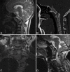
Craniometric measurements (A) Solid black: ADInormal is <3 mm adults and<5 mm children, Dotted black: PADI normal is >13 mm Dotted white: Chamberlain's line and solid white: Mc Rae Line (B) White: Wecken-Heim Clivus base line: It should fall tangent to the posterior aspect of the tip of the odontoid. It forms Clivus Canal angle along the posterior surface of the axis body
Figure 2 (A-C)
Figure 2 (A-C)
Coronal (A) and axial (B) T2W image showing major primary stabilising and sagittal T2W image (C) showing the various secondary stabilising ligaments in relation to odontoid and CVJ. TAL = Transverse atlantal ligament, 1: Anterior atlanto-occipital membrane, 2: ALL, 3: Tectorial membrame, 4: Posterior atlanto-occipital membrane, 5: Ligamentum flavum, 6: PLL
Figure 3 (A-D)
Figure 3 (A-D)
AAD in two different patients Chiari 1 malformation: Sagittal T1W (A) and CT (B) image showing descent of cerebellar tonsil beyond foramen magnum into the cervical canal. Associated basilar invagination, atlanto-axial dislocation and occipitalisation of atlas is seen Klippel feil syndrme: Coronal (C) Sagittal T2W (D) MRI images in a patient with short neck showing multiple segmentation anomalies with AA dislocation occipitalisation of atlas, and chiari I malformation. Associated syrinx (arrow) is also noted
Figure 4 (A and B)
Figure 4 (A and B)
Sagittal CT images (A) and (B) showing Group A and Group B Basilar Invagination respectively. Dotted- Chamberlain's line; Solid- Mc Rae Line
Figure 5 (A and B)
Figure 5 (A and B)
Neural Central Synchondrosis: Coronal (A) and Sagittal (B) reformatted CT images showing ossification of dens in 4 year old child. The tip of basal dental segment is bicornuate from bilateral secondary ossification centres. Lucent gap (arrow) marks synchondrosis which fuses normally fuses with rest of the C2 by 8 years of age. Note terminal ossicle has noted appeared yet
Figure 6 (A-C)
Figure 6 (A-C)
Odontoid aplasia: Sagittal T1W (A) and coronal (B) and sagittal T2W (C) MR images of 28 year old patient with aplastic dens. Excavation defect is seen in the body of odontoid (arrow) below the level of articular process on coronal image. Additional findings include platybasia, short clivus and syrinx (solid arrow) involving the cervical spinal cord
Figure 7 (A and B)
Figure 7 (A and B)
Os odontoideum: Sagittal CT image (A) T1W MRI (B) in a 17 year old patient with quadriparesis showing rounded bony fragment lying above and anterior to the base of dens. Dens is hypoplastic, smooth and well corticated and anterior arch is hypertrophied (arrow) and rounded differentiating the condition from fracture. MR also revealed marked ligament thickening, spinal canal narrowing with cord compression and myelomalacic changes
Figure 8 (A and B)
Figure 8 (A and B)
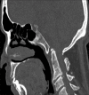
Type II fracture (A) Sagittal CT in a 56 year old patient with trauma showing the fracture fragment that match with the expected defect, the upper margins of axis are sharp and irregular (solid arrow) with normal half moon shaped anterior arch. Condylus Tertius (B) Sagittal CT image in a 39 year old female patient showing an accessory ossification centre (dotted arrow) is seen forming a pseudo-articulation with anterior arch of atlas. The distal most end of clivus is blunted while dens is normal. Note occipitalisation of posterior arch atlas and BI
Figure 9
Figure 9
Persistent os-terminale: The terminal ossicle is seen separate from dens due to failure of fusion. It may be confused with a type 1 odontoid fracture. The odontoid process is usually normal in height
Figure 10 (A and B)
Figure 10 (A and B)
Dens bicornis: Reformatted coronal and Sagittal CT (A and B) images in a 19 year old asymptomatic boy showing bifid tip of the dens. Note that lower synchondrosis has also closed and terminal ossicle has not appeared yet. Note that dens pivot is of normal height
Figure 11
Figure 11
Anderson and D’Alonzo classification of three types of odontoid fractures (#) Type I: Oblique avulsion # through the upper part of the odontoid process at the point of alar ligament attachment. Type II: # occur at the junction of the odontoid process and the body of axis. Type III: # line extend down in to the body of axis
Figure 12 (A and B)
Figure 12 (A and B)
Type II fracture Odontoid: Sagittal T2W (A) and reformatted sagittal CT (B) of 58 year old male showing type 2 odontoid fracture with retropulsion of proximal segment posteriorly. There is injury to posterior longitudinal ligament, cord compression with marrow edema
Figure 13 (A-C)
Figure 13 (A-C)
The axis ring sign: Lateral cervical radiograph of a normal patient (A) Showing an intact axis ring (solid arrow). Plain radiograph (B) and corresponding sagittal CT (C) Image showing disruption of roof of the ring (dotted arrow) in a patient with road trauma suggestive of type III fracture. The ring remains intact in type II fracture
Figure 14 (A-D)
Figure 14 (A-D)
Rotatory atlantoaxial subluxation: A child with h/o trauma followed by torticollis. VRT (A) and coronal and axial CT images showing dens lying eccentrically (B and C) in atlantal ring with anterior subluxation of lateral mass of C1 over C2 (D) Also note that AP axis of C1 and C2 are no more parallel
Figure 15 (A-C)
Figure 15 (A-C)
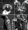
Sagittal CT (A) Sagittal T1W and T2W MRI (B and C) Showing secondary degenerative changes in a patient with multiple segmentation anomalies. Reduced ADI (solid arrow) with sclerosis and osteophtye formation is seen. Fluid signal is noted in predental space (arrow) which normally contains fat/soft tissue. Also seen is Group B Basilar invagination
Figure 16 (A-D)
Figure 16 (A-D)
Tuberculosis: Sagittal reformatted CT (A and B) Images show areas of osseous destruction involving the axis and C3 with associated prevertebral collection. Sagittal T2W (C) and sagittal post contrast T1W (D) MR images show rim enhancing prevertebral collection with epidural extension causing cord compression
Figure 17 (A-C)
Figure 17 (A-C)
Rheumatoid Arthritis: SagittalT2 (A) and T1WMRI (B) Images showing T2 hypointense and T1 isointense pannus (arrows) and thickened TAL. There is destruction of odontoid and compression of cervicomedullary junction. Reformatted sagittal CT image (C) Showing Hook shaped odontoid in another patient with RA
Figure 18 (A-D)
Figure 18 (A-D)
Ankylosing Spondylitis: Reformatted Sagittal CT (A and B) Axial CT (C) and coronal pelvis CT (D) in a 34 year old HLA-B 27 positive male reveals atlanto-axial dislocation, ligament calcification (black arrow), fusion of zygoapophyseal joints (white arrow) and secondary atlanto-ocipital assimilation with bilateral sacroiliac and hip joint ankylosis
Figure 19 (A-D)
Figure 19 (A-D)
CCPD crystal deposition: Reformatted CT images in all three planes (A-C) in a 64 year old female showing calcification of ligaments (solid arrow), type II pathological fracture of odontoid with erosions, sclerosis, osteophytes and narrowing of atlanto-dental interval. Hypointense retro-odontoid mass is seen on T2W MRI (D) images (dotted arrow) along with associated cord compression
Figure 20 (A and B)
Figure 20 (A and B)
Multiple Myeloma: Axial and reformatted sagittal CT images (A and B) Showing well defined punched out lytic lesion involving lower part of odontoid. Multiple other well defined lytic bony lesions were present in skull and spine

References

    1. Hensinger RN, Fielding JW, Hawkins RJ. Congenital anomalies of the odontoid process. Orthop Clin North Am. 1978;9:901–12. - PubMed
    1. Erbengi A, Oge HK. Congenital malformations of the craniovertebral junction: Classification and surgical treatment. Acta Neurochir (Wien) 1994;127:180–5. - PubMed
    1. Redlund-Johnell I, Pettersson H. Radiographic measurements of thecranio-vertebral region. Designed for evaluation of abnormalities in rheumatoidarthritis. Acta Radiol Diagn (Stockh) 1984;25:23–8. - PubMed
    1. McRae DL. Bony abnormalities in the region of the foramen magnum: Correlation of the anatomic and neurologic findings. Acta Radiol. 1953;40:335–54. - PubMed
    1. Chamberlain WE. Basilar impression (Platybasia): A bizarre developmentalanomaly of the occipital bone and upper cervical spine with striking andmisleading neurologic manifestations. Yale J Biol Med. 1939;11:487–96. - PMC - PubMed