Case Reports
. 2016 Jan-Mar;26(1):140-4.
doi: 10.4103/0971-3026.178365.

A case report on paraneoplastic encephalitis associated with astrocytoma - An unknown entity

Affiliations
Case Reports

A case report on paraneoplastic encephalitis associated with astrocytoma - An unknown entity

Yogeshwari S Deshmukh et al. Indian J Radiol Imaging. 2016 Jan-Mar.

Abstract

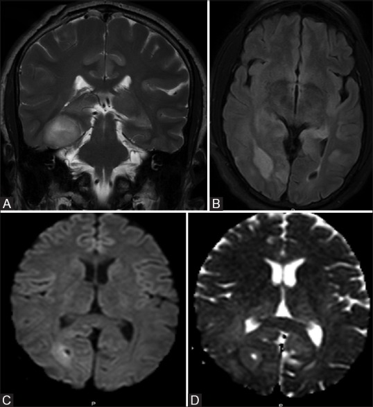
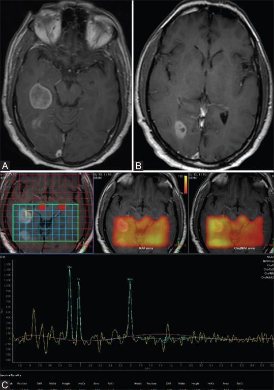
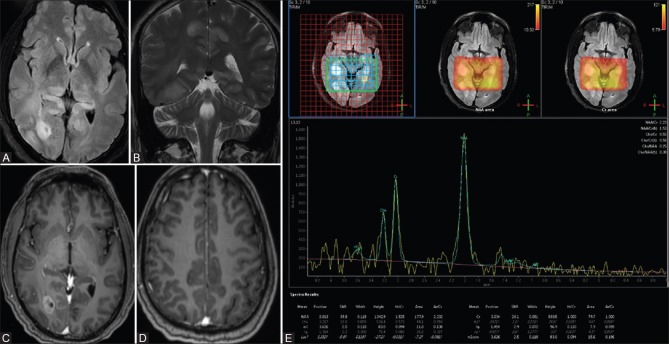
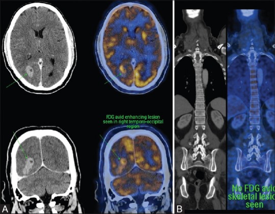
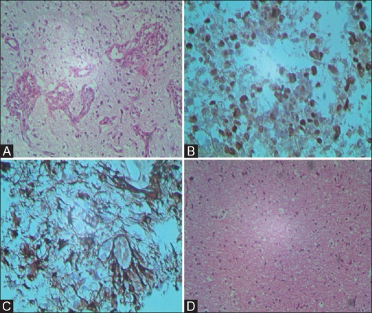
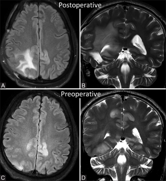
Paraneoplastic encephalitis is a multifocal inflammatory disorder of the central nervous system (CNS) that is associated with remote neoplasias. The most common malignancy associated with it is bronchial carcinoma, typically small cell carcinoma of lung. It has never been described in association with intracranial neoplasm. We present and discuss the clinical, radiological, and histopathological findings of paraneoplastic encephalitis with intracranial space-occupying lesions (SOLs) in a 55-year-old man. He was thoroughly investigated and biopsy revealed presence of astrocytoma with changes of paraneoplastic encephalitis.

Keywords: Astrocytoma; limbic encephalitis; paraneoplastic encephalitis.


Figures

Figure 1 (A-D)
Figure 1 (A-D)
FLAIR axial image showing two well-defined focal mass lesions in the right temporal and occipital lobes (A and B). DWI showing restriction of diffusion in the wall of the lesions in the right temporal/occipital lobe (C and D)
Figure 2 (A-C)
Figure 2 (A-C)
(A-C) Post-contrast T1 sagittal image shows heterogeneous enhancement of lesions in the right temporal and occipital lobes (A and B). Multivoxel intermediate TE MR spectroscopywith mild elevation of choline in mass lesions, with choline to creatine ratio range is 1.3-1.9 (C)
Figure 3 (A-E)
Figure 3 (A-E)
FLAIR and T2W images show multiple focal hyperintensities in bilateral hippocampi, dorsal thalami, and cingulate gyri (A and B), no enhancement was seen on post-contrast study in hippocampi and affected cortices (C and D). Multivoxel intermediate TE MR spectroscopy shows normal choline peak (E)
Figure 4 (A and B)
Figure 4 (A and B)
PET CT showing avid FDG uptake in intracranial masses and no abnormal FDG avid activity in the rest of the body (A and B)
Figure 5 (A-D)
Figure 5 (A-D)
200× H and E and immunohistochemistry slide shows multiple neoplastic astrocytes with high mitotic index (A-D). 200× H and E slide shows reactive gliosis with interstitial and perivascular infiltrates of lymphocytes, a feature of paraneoplastic encephalitis (D)
Figure 6 (A-D)
Figure 6 (A-D)
FLAIR axial and T2W coronal images in follow-up MRI done after 6 months reveal significant decrease in cortical hyperintensity in bilateral cigulate gyri when compared with preoperative initial MRI images (C and D)

References

    1. Dalmau J, Rosenfeld MR. ParaneoplasticsyndromesoftheCNS. Lancet Neurol. 2008;7:327–40. - PMC - PubMed
    1. Antoine JC, Honnorat J, Anterion CT, Aguera M, Absi L, Fournel P, et al. Limbic encephalitis and immunological perturbations in two patients with thymoma. J NeurolNeurosurg Psychiatry. 1995;58:706–10. - PMC - PubMed
    1. Grisold W, Giometto B, Vitaliani R, Oberndorfer S. Current approaches to the treatment of paraneoplastic encephalitis. TherAdvNeurolDisord. 2011;4:237–48. - PMC - PubMed
    1. Graus F, Vincent A, Pozo-Rosich P, Sabater L, Saiz A, Lang B, et al. Anti-glial nuclear antibody: Marker of lung cancer-related paraneoplastic neurological syndromes. J Neuroimmunol. 2005;165:166–71. - PMC - PubMed
    1. Seki M, Suzuki S, Ishii K, Izawa Y, Takahashi S, Toyama Y, et al. Differential diagnosis between intracranial dissemination of spinal cord astrocytoma and paraneoplastic limbic encephalitis. Intern Med. 2012;51:321–4. - PubMed

Publication types