. 2016 Apr 14:8:38.
doi: 10.1186/s13148-016-0205-6. eCollection 2016.

The epigenetic modifier JMJD6 is amplified in mammary tumors and cooperates with c-Myc to enhance cellular transformation, tumor progression, and metastasis

Affiliations

The epigenetic modifier JMJD6 is amplified in mammary tumors and cooperates with c-Myc to enhance cellular transformation, tumor progression, and metastasis

Olga Aprelikova et al. Clin Epigenetics. .

Abstract

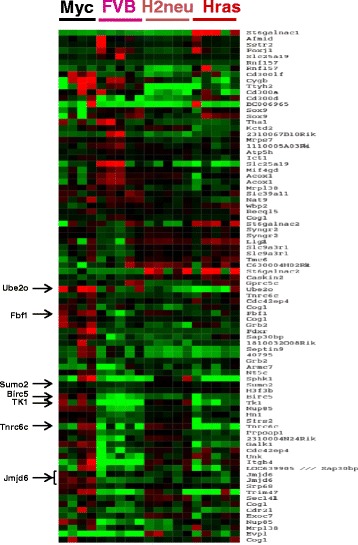
Background: Oncogene overexpression in primary cells often triggers the induction of a cellular safeguard response promoting senescence or apoptosis. Secondary cooperating genetic events are generally required for oncogene-induced tumorigenesis to overcome these biologic obstacles. We employed comparative genomic hybridization for eight genetically engineered mouse models of mammary cancer to identify loci that might harbor genes that enhance oncogene-induced tumorigenesis.

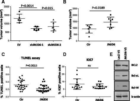
Results: Unlike many other mammary tumor models, the MMTV-Myc tumors displayed few copy number variants except for amplification of distal mouse chromosome 11 in 80 % of the tumors (syntenic to human 17q23-qter often amplified in human breast cancer). Analyses of candidate genes located in this region identified JMJD6 as an epigenetic regulatory gene that cooperates with Myc to enhance tumorigenesis. It suppresses Myc-induced apoptosis under varying stress conditions through inhibition of p19ARF messenger RNA (mRNA) and protein, leading to reduced levels of p53. JMJD6 binds to the p19ARF promoter and exerts its inhibitory function through demethylation of H4R3me2a. JMJD6 overexpression in MMTV-Myc cell lines increases tumor burden, induces EMT, and greatly enhances tumor metastasis. Importantly, we demonstrate that co-expression of high levels of JMJD6 and Myc is associated with poor prognosis for human ER+ breast cancer patients.

Conclusions: A novel epigenetic mechanism has been identified for how JMJD6 cooperates with Myc during oncogenic transformation. Combined high expression of Myc and JMJD6 confers a more aggressive phenotype in mouse and human tumors. Given the pleiotropic pro-tumorigenic activities of JMJD6, it may be useful as a prognostic factor and a therapeutic target for Myc-driven mammary tumorigenesis.

Keywords: Copy number variants; Epigenetics; JMJD6; Mammary cancer; Myc; Tumor progression.


Figures

Fig. 1
Fig. 1
DNA copy number analysis. Array CGH analysis of mouse mammary gland tumors from eight genetically engineered models of breast cancer. 5–6 tumors were used for each model. The threshold line is drawn at 35 % of samples. Genomic regions of significant gains are shown in blue and significant losses are shown in red. Arrow indicates the amplification of distal mouse chromosome 11
Fig. 2
Fig. 2
Gene expression microarray analysis for chromosome 11 amplified region. The heatmap shows the differential gene expression in mammary gland tumors from MMTV-Myc transgenic mice with chromosome 11 amplification versus MMTV-Her2, or MMTV-HRas tumors lacking the chromosome 11 amplification, or normal lactating mammary glands from FVB/N mice. Genes labeled in red are expressed at higher than median levels and genes labeled in green are expressed at lower than median levels. Genes selected for further validation are indicated on the left side
Fig. 3
Fig. 3
Analysis of cell death induced by glucose deprivation or etoposide treatment in cells with JMJD6 knock-down. a Myc83-derived cell lines (parental line from a MMTV-Myc tumor) with stable expression of two independent shRNAs targeting JMJD6 or with empty vector (EV) control were treated with 100 μM etoposide or grown in glucose-free media for 20 h. Cell death was measured using CytoTox-Glo reagent. The experiments were repeated 3 times and the percentage of dead cells in each experiment was expressed relative to control (EV) cells. b NMuMG cells with MycER™ and shJMJD6 expression were first treated with 150 nM 4-OHT (MycON) or ethanol (MycOFF) for 24 h to activate Myc and then treated as in a Black bars—MycOFF (ethanol-treated cells), open bars—MycON (4-OHT-treated cells). Results were normalized to EV control with MycOFF. *p < 0.05, **p < 0.01
Fig. 4
Fig. 4
Overexpression of wild-type, but not mutated, JMJD6 suppresses Myc-induced cell death in response to different stress conditions. NMuMG cells stably expressing MycERTM or empty pBabe vector were transduced with JMJD6-V5 or LacZ-V5 control. Cells were treated with 4-OHT or ethanol as in Fig. 3 and placed in glucose-free (a), serum-free (b), or glutamine-free (c) media or treated with etoposide (d) for another 20 h. Cell death was measured as in Fig. 3. Black bars—MycOFF, open bars—MycON. e Catalytically inactive JMJD6H187A (JMJD6mut) is not able to suppress Myc-induced cell death. *p < 0.05, **p < 0.01. f Western blot analysis shows equal ectopic expression of JMJD6 or mutated JMJD6 in NMuMG cells with our without Myc expression
Fig. 5
Fig. 5
Western blot analysis of apoptotic markers in Myc-induced cells in the presence of wild-type or mutated JMJD6. NMuMG cells were treated with 150 nM 4-OHT to induce Myc or ethanol and placed in glucose- or glutamine-free media or treated with 100 mM of etoposide. Expression of cleaved PARP and cleaved caspase 3 was determined by Western blot demonstrating reduced levels in the presence of wild-type, but not mutant, JMJD6
Fig. 6
Fig. 6
JMJD6 overexpression reduces p53 and p19ARF levels. a Immunoblotting of NMuMG cells with ectopic expression of JMJD6 or LacZ control shows decreased protein levels of p53 and p19ARF in cells with or without Myc induction. b Cells were stimulated with 4-OHT to induce Myc and then treated with 100 mM etoposide for 4 and 8 h to analyze p53 induction. Immunoblots show lower levels of p53 or Ser18-phosphorylated p53 as well as p19ARF and p21 in cells expressing JMJD6. Intensity of each band normalized to β-actin was obtained using ImageJ software. c Overexpression of p19ARF in the presence of JMJD6 restores Myc-induced cell death. NMuMG cells expressing MycER™ together with JMJD6, LacZ, or JMJD6 plus p19ARF were treated with 4-OHT (MycON) or ethanol (MycOFF) for 24 h and placed in serum-free media for another 24 h. Percentage of dead cells was measured with CytoTox-Glo reagent. Black bars—MycON, open bars—MycOFF. d Western blot analysis of NMuMG cells with ectopic overexpression of p19ARF. e RT-qPCR analysis shows that overexpression of JMJD6 in NMuMG cells inhibits p19ARF transcription. Black bars—NMuMG cells expressing LacZ control, open bars—cells expressing wild-type JMJD6. f Knock-down of endogenous JMJD6 with three different shRNAs increases mRNA levels of p19ARF
Fig. 7
Fig. 7
JMJD6 binds to the p19ARF promoter and decreases histone H4R3 asymmetric dimethylation of the p19ARF promoter. ChIP analysis of the p19ARF promoter in NMuMG cells expressing LacZ, JMJD6, or JMJD6 mutant using immunoprecipitation with a anti-H4R4me2a, b anti-JMJD6 antibody, c V5-conjugated agarose. d ChIP analysis of HeLa cells shows binding of endogenous JMJD6 to the human p14ARF promoter. Two different primer sets (distal and proximal) were used for qPCR after immunoprecipitation. Primers for human β-globin gene promoter were used as a positive control
Fig. 8
Fig. 8
JMJD6 promotes tumor growth of cells derived from MMTV-Myc mammary gland tumors. a 106 Myc83 cells (with amplification of the chromosome 11 locus containing JMJD6) stably expressing empty vector (EV) or two independent shRNAs targeting JMJD6 were injected into mammary fat pads of FVB/N mice and tumors were measured over the next month. b 5 × 105 88CT1 cells (no chromosome 11 amplification) with stable expression of LacZ control or wild-type JMJD6 were surgically implanted into mammary fat pads and tumor growth was measured over the next 20 days. c Tumors from b were formalin fixed and sectioned and the percentage of dead cells was analyzed by TUNEL assay. d Tumors from b were immunostained with anti-Ki67 antibodies and signal intensity was quantitated by ImageJ software. e Western blot analysis of pro-survival genes in 88CT1 cells expressing JMJD6
Fig. 9
Fig. 9
JMJD6 increases the metastatic propensity of c-Myc-expressing cells. Transwell migration (a) and invasion (b) assays of 88CT1 cells overexpressing JMJD6 compared to LacZ control (*p < 0.05, **p < 0.01, respectively). c Western blot analysis of EMT markers in the cells used in a and b shows increased expression of Snail and Twist1 in cells with high expression of JMJD6. d Lung colonization in vivo. 5 × 105 cells used in a and b were injected by tail vein into FVB/N mice. After 20 days, the lungs were formalin fixed and stained with H&E. e Quantitation of metastatic nodules per lung of mice shown in d
Fig. 10
Fig. 10
High expression of JMJD6 and Myc shows the worst prognosis for human mammary gland tumors. a Kaplan-Meier survival curves are shown for the high versus low expression of JMJD6 in the presence of low (left panel) or high (right panel) expression of Myc. METABRIC database (~2000 patients) was used to perform this analysis. Analysis was performed as in a for b ER-negative or c ER-positive mammary gland tumors

References

    1. Green JE, Shibata MA, Yoshidome K, Liu ML, Jorcyk C, Anver MR, Wigginton J, Wiltrout R, Shibata E, Kaczmarczyk S, et al. The C3(1)/SV40 T-antigen transgenic mouse model of mammary cancer: ductal epithelial cell targeting with multistage progression to carcinoma. Oncogene. 2000;19:1020–7. doi: 10.1038/sj.onc.1203280. - DOI - PubMed
    1. Hennighausen L. Mouse models for breast cancer. Oncogene. 2000;19:966–7. doi: 10.1038/sj.onc.1203346. - DOI - PubMed
    1. Herschkowitz JI, Simin K, Weigman VJ, Mikaelian I, Usary J, Hu Z, Rasmussen KE, Jones LP, Assefnia S, Chandrasekharan S, et al. Identification of conserved gene expression features between murine mammary carcinoma models and human breast tumors. Genome Biol. 2007;8:R76. doi: 10.1186/gb-2007-8-5-r76. - DOI - PMC - PubMed
    1. Foulkes WD, Stefansson IM, Chappuis PO, Begin LR, Goffin JR, Wong N, Trudel M, Akslen LA. Germline BRCA1 mutations and a basal epithelial phenotype in breast cancer. J Natl Cancer Inst. 2003;95:1482–5. doi: 10.1093/jnci/djg050. - DOI - PubMed
    1. Sorlie T, Tibshirani R, Parker J, Hastie T, Marron JS, Nobel A, Deng S, Johnsen H, Pesich R, Geisler S, et al. Repeated observation of breast tumor subtypes in independent gene expression data sets. Proc Natl Acad Sci U S A. 2003;100:8418–23. doi: 10.1073/pnas.0932692100. - DOI - PMC - PubMed

Publication types

MeSH terms

Substances