. 2016 Apr 15;11(4):e0153632.
doi: 10.1371/journal.pone.0153632. eCollection 2016.

Decreased Polycystin 2 Levels Result in Non-Renal Cardiac Dysfunction with Aging

Affiliations

Decreased Polycystin 2 Levels Result in Non-Renal Cardiac Dysfunction with Aging

Ivana Y Kuo et al. PLoS One. .

Abstract

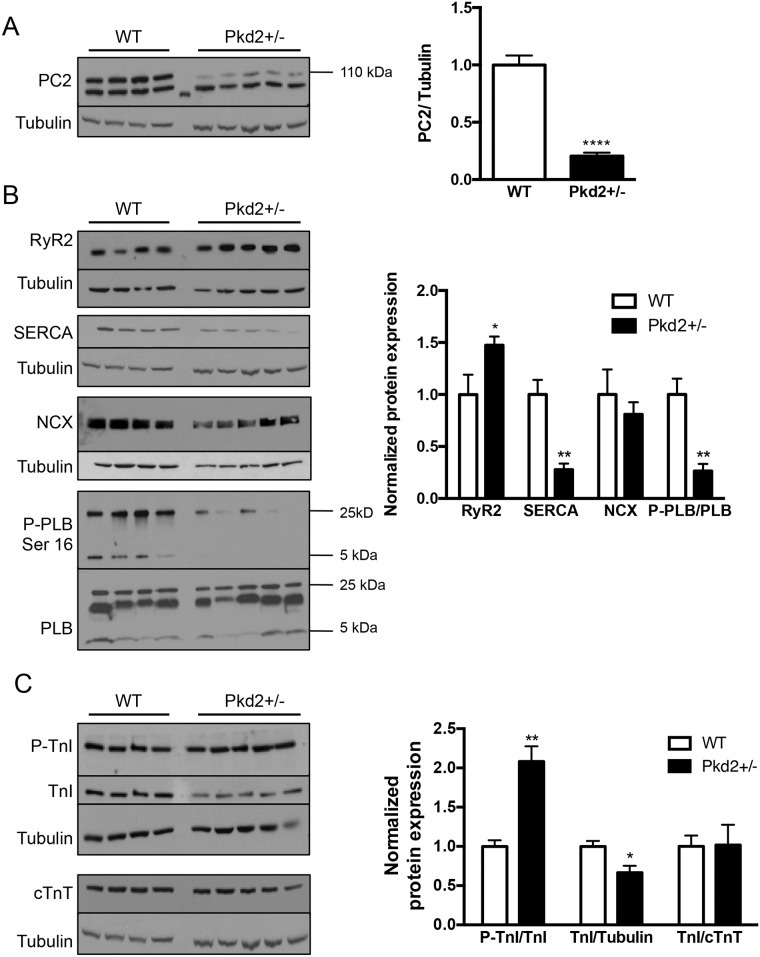
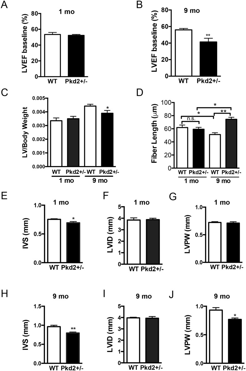
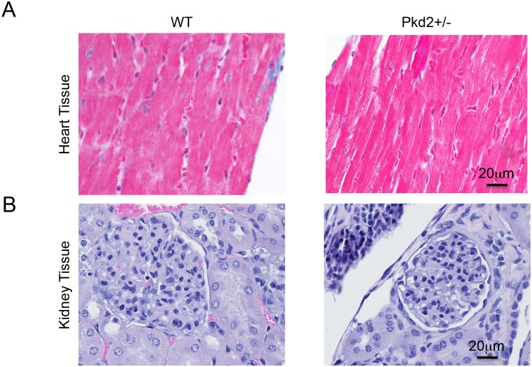
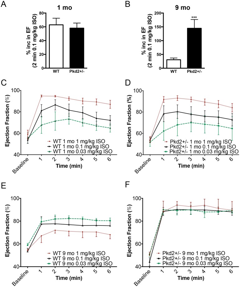
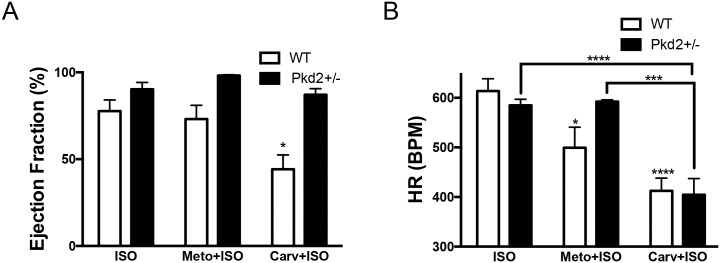
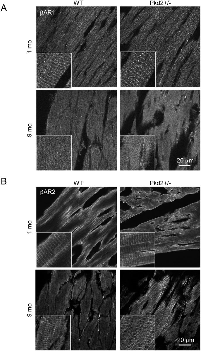
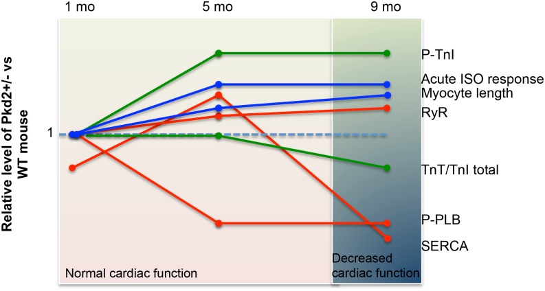
Mutations in the gene for polycystin 2 (Pkd2) lead to polycystic kidney disease, however the main cause of mortality in humans is cardiac related. We previously showed that 5 month old Pkd2+/- mice have altered calcium-contractile activity in cardiomyocytes, but have preserved cardiac function. Here, we examined 1 and 9 month old Pkd2+/- mice to determine if decreased amounts of functional polycystin 2 leads to impaired cardiac function with aging. We observed changes in calcium handling proteins in 1 month old Pkd2+/- mice, and these changes were exacerbated in 9 month old Pkd2+/- mice. Anatomically, the 9 month old Pkd2+/- mice had thinner left ventricular walls, consistent with dilated cardiomyopathy, and the left ventricular ejection fraction was decreased. Intriguingly, in response to acute isoproterenol stimulation to examine β-adrenergic responses, the 9 month old Pkd2+/- mice exhibited a stronger contractile response, which also coincided with preserved localization of the β2 adrenergic receptor. Importantly, the Pkd2+/- mice did not have any renal impairment. We conclude that the cardiac-related impact of decreased polycystin 2 progresses over time towards cardiac dysfunction and altered adrenergic signaling. These results provide further evidence that polycystin 2 provides a critical function in the heart, independent of renal involvement.


Conflict of interest statement

Competing Interests: The authors have declared that no competing interests exist.

Figures

Fig 1
Fig 1. Expression of calcium handling and contractile proteins in 9 mo WT and Pkd2+/- mice.
(A) PC2 expression in the left ventricle was diminished by ~70% in Pkd2+/- mice compared with WT mice. (B) Expression of the intracellular calcium release channel RyR2 was increased, whereas SERCA2A and the phosphorylated form of PLB were decreased. NCX levels were unchanged. Data are quantified on the right. RyR2, SERCA2A and NCX are all normalized to tubulin. (C) The expression of the phosphorylated form of TnI was significantly increased in the Pkd2+/- mice. The levels of TnI and cTnT in the Pkd2+/- mice were diminished overall compared to the WT mice. Data are quantified on the right. Each lane represents a separate animal. *p < 0.05, **p < 0.01, ****p<0.0001.
Fig 2
Fig 2. 9 mo Pkd2+/- mice have decreased ejection fraction and ventricular remodeling.
(A) 1 mo WT and Pkd2+/- mice have similar left ventricular ejection fraction values. (B) 9 mo Pkd2+/- mice have significantly lower left ventricular ejection fraction values compared with WT mice. (C) Left ventricular to body weight ratios were significantly reduced in 9 mo mice but were unchanged in 1 mo mice. (D) Cardiomyocyte lengths were significantly longer in 9 mo mice but are unchanged in 1 mo mice. (E-G) 1 mo Pkd2+/- mice have significantly thinner inner septum measurements compared with WT mice, but the posterior wall and interior diameter are the same. (H-J) 9 mo Pkd2+/- mice have significantly thinner left ventricular wall and inner septum measurements compared with WT mice. Data in A-C, E-J are representative of five 1 mo mice in each group, and eight and nine WT and Pkd2+/- 9 mo mice, respectively. Data in D are representative of at least two different animals per group with 22–45 cells measured from each animal. *p < 0.05, **p < 0.01, n.s. = not significant.
Fig 3
Fig 3. No gross cardiac or renal abnormalities in WT and Pkd2+/- mice.
(A) Left ventricles from 9 mo WT (left) and Pkd2+/- (right) mice show similar Masson’s Trichrome staining patterns with no signs of fibrosis. (B) 9 mo WT (left) and Pkd2+/- (right) mice have similar H&E staining patterns with no renal cysts. Data are representative of at least 3 mice in each group.
Fig 4
Fig 4. 9 mo Pkd2+/- mice have enhanced responses to acute ISO application.
(A, B) 1 mo WT and Pkd2+/- mice have comparable left ventricular ejection fraction response 2 min after acute 0.1 mg/kg ISO injection (A) whereas 9 mo Pkd2+/- have a significantly enhanced response (B). (C,D) Using three doses of ISO (0.03, 0.1 and 1 mg/kg), 1 mo WT (C) and Pkd2+/- (D) mice have similar left ventricular ejection fraction responses. (E,F) Using three doses of ISO (0.03, 0.1 and 1 mg/kg), 9 mo WT mice (E) have a dose dependent response, though blunted compared to the 1 mo mice (left). 9 mo Pkd2+/- mice (F) have maximal left ventricular ejection fraction responses to ISO, regardless of the dose. For 1 mo mice, data are representative of 5 mice for each genotype for the 0.1 mg/kg and 1 mg/kg data. For 9 mo mice, the 0.1 mg/kg ISO data are representative of 8 and 9 WT and Pkd2+/-, respectively. For the 0.03 mg/kg ISO data, the 1 mo and 9 mo data are representative of 3 mice. The 1 mg/kg ISO data for 9 mo mice are representative of 3 mice per group.
Fig 5
Fig 5. 9 mo Pkd2+/- mice have blunted responses to β-blockers.
(A) Pre-treating WT and Pkd2+/- mice with the βAR-1 blocker metoprolol does not alter the ejection fraction after ISO challenge, whereas pre-treatment with the pan β-blocker carvedilol diminishes the WT response but not the Pkd2+/- response. (B) The heart rate was significantly decreased after either metoprolol or carvedilol pre-treatment followed by ISO challenge in WT mice, compared to ISO only challenge. In Pkd2+/- mice, only carvedilol, not metoprolol has an effect on heart rate. Note that heart rate after carvedilol pre-treatment is the not significantly different in WT and Pkd2+/- mice. The data are representative of 3–4 mice per group.
Fig 6
Fig 6. β-Adrenergic receptor expression is altered in 9 mo Pkd2+/- mice.
(A) Staining intensity for βAR-1 was similar in 1 and 9 mo WT and Pkd2+/- mice. (B) Staining intensity for βAR-2 was similar in 1 mo WT and Pkd2+/- mice, but increased in 9 mo Pkd2+/- mice, and had a more striated pattern in the Pkd2+/- mice. Insets represent higher magnification images. Images are representative of staining taken from at least 3 separate mice.
Fig 7
Fig 7. Summary of the changes in protein expression and cardiac function in the Pkd2+/- mice examined over a 9 mo period.
Data is summarized from the current paper as well as data from 5 mo mice [15]. Each line represents the expression levels of various proteins (SERCA, RyR, p-TnI, TnI, p-PLB) or other functional readouts (eg: cardiomyocyte length, acute response to ISO) at 1, 5 and 9 mo of age. The orange lines represent those associated with the calcium signaling apparatus (SERCA, RyR, PLB). The green lines represents those proteins associated with the contractile apparatus (p-TnI and cTnT). The blue lines represent other measurements (cardiomyocyte cell length and response to acute ISO stimulus). The background shading (pink and blue) represents the overall cardiac function of the mouse, as measured with echocardiogram.

References

    1. Reeders ST. Multilocus polycystic disease. Nat Genet. 1992;1(4):235–7. 10.1038/ng0792-235 . - DOI - PubMed
    1. Helal I, Reed B, Mettler P, Mc Fann K, Tkachenko O, Yan XD, et al. Prevalence of cardiovascular events in patients with autosomal dominant polycystic kidney disease. Am J Nephrol. 2012;36(4):362–70. Epub 2012/10/06. 10.1159/000343281 . - DOI - PMC - PubMed
    1. Schrier RW. Renal volume, renin-angiotensin-aldosterone system, hypertension, and left ventricular hypertrophy in patients with autosomal dominant polycystic kidney disease. J Am Soc Nephrol. 2009;20(9):1888–93. Epub 2009/08/22. ASN.2008080882 [pii] 10.1681/ASN.2008080882 . - DOI - PubMed
    1. Ecder T, Schrier RW. Cardiovascular abnormalities in autosomal-dominant polycystic kidney disease. Nature reviews Nephrology. 2009;5(4):221–8. Epub 2009/03/27. 10.1038/nrneph.2009.13 - DOI - PMC - PubMed
    1. Torres VE, Abebe KZ, Chapman AB, Schrier RW, Braun WE, Steinman TI, et al. Angiotensin blockade in late autosomal dominant polycystic kidney disease. N Engl J Med. 2014;371(24):2267–76. 10.1056/NEJMoa1402686 - DOI - PMC - PubMed

Publication types

MeSH terms