. 2016 May;89(5):1027-1036.
doi: 10.1016/j.kint.2015.12.046. Epub 2016 Mar 9.

Osteopontin protects against high phosphate-induced nephrocalcinosis and vascular calcification

Affiliations

Osteopontin protects against high phosphate-induced nephrocalcinosis and vascular calcification

Neil J Paloian et al. Kidney Int. 2016 May.

Abstract

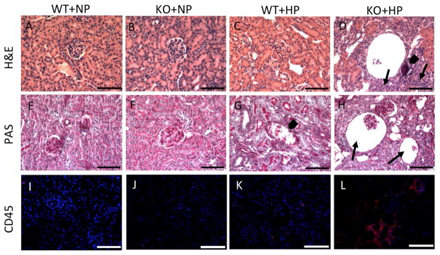
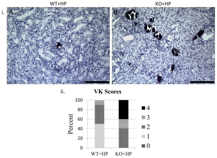
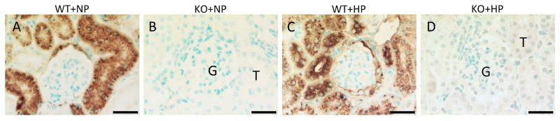
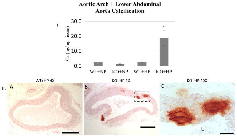
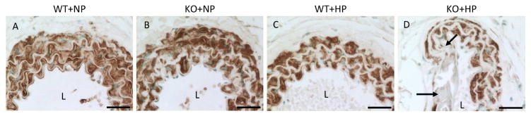
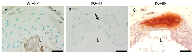
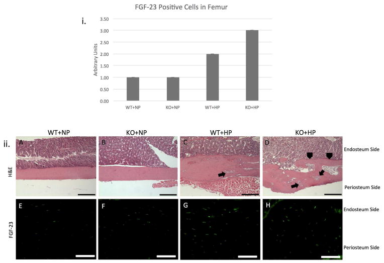
Pathologic calcification is a significant cause of increased morbidity and mortality in patients with chronic kidney disease. The precise mechanisms of ectopic calcification are not fully elucidated, but it is known to be caused by an imbalance of procalcific and anticalcific factors. In the chronic kidney disease population, an elevated phosphate burden is both highly prevalent and a known risk factor for ectopic calcification. Here we tested whether osteopontin, an inhibitor of calcification, protects against high phosphate load-induced nephrocalcinosis and vascular calcification. Osteopontin knockout mice were placed on a high phosphate diet for 11 weeks. Osteopontin deficiency together with phosphate overload caused uremia, nephrocalcinosis characterized by substantial renal tubular and interstitial calcium deposition, and marked vascular calcification when compared with control mice. Although the osteopontin-deficient mice did not exhibit hypercalcemia or hyperphosphatemia, they did show abnormalities in the mineral metabolism hormone fibroblast growth factor-23. Thus, endogenous osteopontin plays a critical role in the prevention of phosphate-induced nephrocalcinosis and vascular calcification in response to high phosphate load. A better understanding of osteopontin's role in phosphate-induced calcification will hopefully lead to better biomarkers and therapies for this disease, especially in patients with chronic kidney disease and other at-risk populations.

Keywords: calcium; fibroblast growth factor-23; parathyroid hormone; phosphate; uremia; vascular calcification.


Conflict of interest statement

Disclosure

All authors have no competing interests.

Figures

Figure 1
Figure 1. Loss of normal renal structure in the KO+HP group
H&E staining of the WT+NP (A), KO+NP (B), and WT+HP (C) kidneys are normal. H&E staining of the kidneys of the KO+HP group (D) demonstrate a marked cellular infiltrate (arrows) surrounding both dilated tubular structures and mineral deposits (D, arrowhead). PAS staining of the WT+NP (E) and KO+NP (F) kidneys is normal. Occasional tubular mineral deposits are seen on PAS staining of WT+HP kidneys (G, arrowhead) with otherwise normal morphology. PAS staining of KO+HP kidneys (H) demonstrates cystic glomeruli (arrows) with marked dilatation of Bowman’s capsule. Dilated tubules are also seen. CD45 IF of the WT+NP (I) and KO+NP (J) indicates the presence of occasional inflammatory cells. The WT+HP group shows mild subcapsular and interstitial inflammation. A significant inflammatory infiltrate is present near cystic, dilated glomeruli and calcification sites (orange in panel L). Original magnification = 20X. Scale bar = 75 μm.
Figure 2
Figure 2. OPN KO mice on HP diet develop abundant renal calcium phosphate deposition
i. VK staining confirms presence of calcium phosphate deposition throughout the kidney of KO+HP mice (B). Occasional renal mineral deposition is seen in the WT+HP mice (A). Original magnification = 10X. Scale bar = 150 μm. ii. VK scoring for mineral granules demonstrates a marked increase in renal calcium deposits in the KO+HP group when compared to the WT+HP group. 0 = no granules found in the section, 1 = 1–25 granules, 2 = 26–75 granules, 3 = 76–150 granules, 4 = >150 granules
Figure 3
Figure 3. OPN Immunohistochemistry confirms depletion of OPN protein in KO kidney
Kidney sections of WT+NP (A) and WT+HP (C) demonstrate abundant renal tubular OPN expression. OPN expression is absent in the KO+NP (B) and KO+HP (D) kidneys. Original magnification = 40X. Scale bar = 30 μm. G = glomerulus; T = Tubule.
Figure 4
Figure 4. KO+HP mice develop vascular calcification
i. Aortic arch plus lower abdominal aorta biochemical calcium content expressed as μg calcium normalized to mg dry weight (mean±s.e.m.). *p<0.05 compared to the WT+NP, KO+NP, and WT+HP. Calcification was not different between the WT+NP, KO+NP, and WT+HP groups. ii. Alizarin Red stains of the abdominal aorta show marked calcification in the KO+HP group (B) and absence of mineral in the WT+HP group (A). Original magnification = 4X (cropped). Scale bar = 350 μm. Magnification of the mineral (box) in the KO+HP aorta (C) shows that the mineral is located entirely in the medial layer of the vessel. Original magnification = 40X. Scale bar = 30 μm. L = lumen.
Figure 5
Figure 5. Loss of sm22 alpha expression in the KO+HP aorta confirms phenotypic transformation of VSMCs
Sm22α expression is normal throughout the length of the abdominal aorta in the WT+NP (A), KO+NP (B), and WT+HP (C) groups.) Sm22α expression is absent in areas of the aorta (arrows) in the KO+HP mice (D) consistent with phenotypic transformation of the VSMC seen in vascular calcification. Original magnification = 40X. Scale bar = 30 μm. L = lumen.
Figure 6
Figure 6. OPN expression is absent in the calcified vasculature of the KO+HP group
OPN IHC of the abdominal aorta of the WT+HP mice (A) demonstrates no OPN expression in the vessel as expected in the absence of calcification. Despite the presence of mineral (arrow) the KO+HP aorta (B) is devoid of OPN. An adjacent section of the KO+HP aorta (C) is stained with Alizarin Red to highlight the mineral. Original magnification = 40X. Scale bar = 30 μm. L = lumen.
Figure 7
Figure 7. FGF-23 expression is increased in OPN+HP femurs
i. FGF-23 expression is elevated in femurs of WT+HP, but expression is highest in KO+HP. ii. H&E staining of femurs demonstrates widening of the diaphysis and increased cortical porosity (arrows) in both HP fed mice (C,D). Large protrusions of trabeculae (arrowheads) are noted exclusively in the KO+HP group (D). There is more new bone forming in the KO + HP group, and therefore more osteocytes expressing FGF23 per bone area as seen in the IF. H&E: Original magnification = 4X. Scale bar = 350uM. FGF-23 IF: Original magnification = 20X. Scale bar = 75uM.

References

    1. Shavit L, Jaeger P, Unwin RJ. What is nephrocalcinosis? Kidney Int. 2015;88:35–43. - PubMed
    1. Witteman JC, Kok FJ, van Saase JL, et al. Aortic calcification as a predictor of cardiovascular mortality. Lancet. 1986;2:1120–1122. - PubMed
    1. Witteman JC, Kannel WB, Wolf PA, et al. Aortic calcified plaques and cardiovascular disease (the Framingham Study) Am J Cardiol. 1990;66:1060–1064. - PubMed
    1. Adragao T, Pires A, Lucas C, et al. A simple vascular calcification score predicts cardiovascular risk in haemodialysis patients. Nephrol Dial Transplant. 2004;19:1480–1488. - PubMed
    1. Russo D, Corrao S, Battaglia Y, et al. Progression of coronary artery calcification and cardiac events in patients with chronic renal disease not receiving dialysis. Kidney Int. 2011;80:112–118. - PMC - PubMed

Publication types

MeSH terms

LinkOut - more resources