Review
. 2016 Jun;39(6):378-393.
doi: 10.1016/j.tins.2016.03.005. Epub 2016 Apr 12.

Emerging Roles for CSF-1 Receptor and its Ligands in the Nervous System

Affiliations
Review

Emerging Roles for CSF-1 Receptor and its Ligands in the Nervous System

Violeta Chitu et al. Trends Neurosci. 2016 Jun.

Abstract

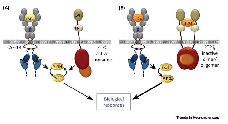
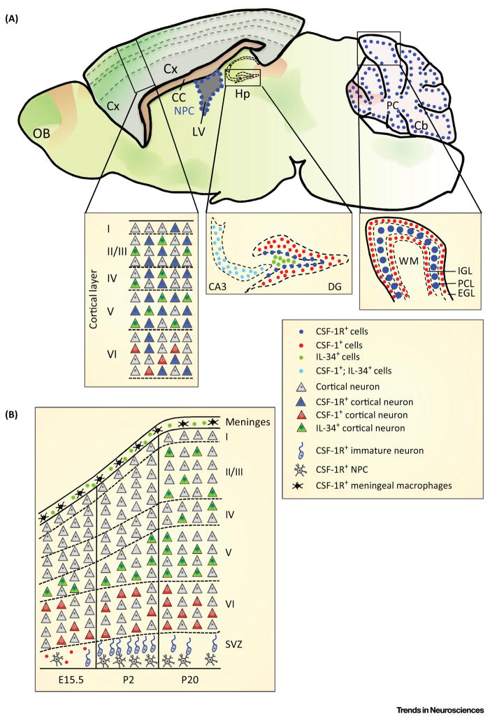
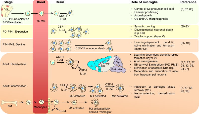
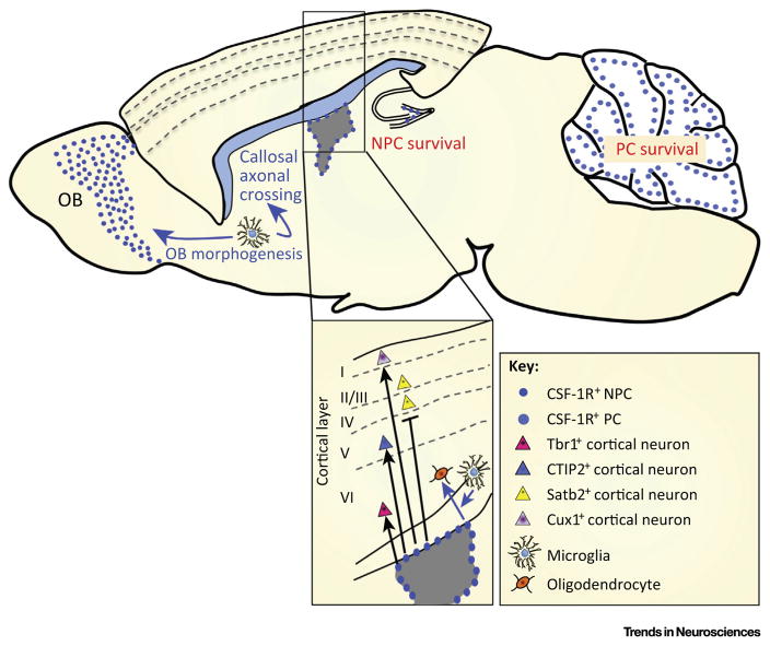
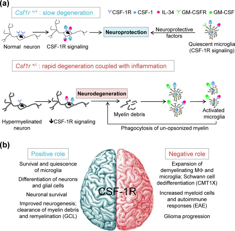
The colony-stimulating factor-1 receptor (CSF-1R) kinase regulates tissue macrophage homeostasis, osteoclastogenesis, and Paneth cell development. However, recent studies in mice have revealed that CSF-1R signaling directly controls the development and maintenance of microglia, and cell autonomously regulates neuronal differentiation and survival. While the CSF-1R-cognate ligands, CSF-1 and interleukin-34 (IL-34) compete for binding to the CSF-1R, they are expressed in a largely non-overlapping manner by mature neurons. The recent identification of a dominantly inherited, adult-onset, progressive dementia associated with inactivating mutations in the CSF-1R highlights the importance of CSF-1R signaling in the brain. We review the roles of the CSF-1R and its ligands in microglial and neural development and function, and their relevance to our understanding of neurodegenerative disease.

Keywords: IL-34; adult-onset leukoencephalopathy with axonal spheroids and pigmented glia; microglia; neural development; neurodegenerative disease; neuronal survival.


Figures

Figure I
Figure I
Putative Mechanism for Differential Regulation by CSF-1 and IL-34 in Cells Coexpressing CSF-1R and PTPζ. (A) CSF-1 signaling: CSF-1 activates the CSF-1R tyrosine kinase leading to increased cellular tyrosine phosphorylation. Catalytically-active PTPζ decreases cellular tyrosine phosphorylation. (B) IL-34 signaling. Like CSF-1, IL-34 activates the CSF-1R, but also binds to PTPζ and inhibits its tyrosine phosphatase activity, further increasing tyrosine phosphorylation and downstream responses, such as NPC differentiation.
Figure 1
Figure 1
Expression of Colony Stimulating Factor-1 Receptor (CSF-1R) and its Ligands in Brain. (A) Expression of CSF-1 (red shading) and interleukin-34 (IL-34) (green shading) in the adult brain (based on Allen Brain Atlas and [–8]). Colocalization studies revealed that, apart from microglia (not shown), CSF-1R (blue) is expressed in ~30% of mature cortical neurons in the forebrain, in hippocampal cells, and in Purkinje neurons. CSF-1 expression (red) is low and restricted to the specific areas of the olfactory bulb, cortex, corpus callosum, hippocampus, and cerebellum. By contrast, IL-34 (green) is expressed throughout the telencephalon but is absent from the cerebellum. With the exception of the CA3 area of the hippocampus, there is no significant overlap in the cellular expression of CSF-1 and IL-34. (B) Dynamics of CSF-1R and ligand expression during cortical development. For clarity, CSF-1R+ microglia, which are present throughout the cortex and stain strongly in the SVZ, are not shown. Abbreviations: Cb, cerebellum; Cx, cortex; CC, corpus callosum; DG, dentate gyrus; EGL, external granular layer; Hp, hippocampus; IGL, internal granular layer; LV, lateral ventricle; NPCs, neural progenitor cells; OB, olfactory bulb; PC, Purkinje cells; PCL, PC layer; SVZ, subventricular zone; WM, white matter.
Figure 2
Figure 2
Contribution of CSF-1R Ligands to the Development, Maintenance, and Activation of Microglia. Primitive microglia progenitors (macrophages) generated in the yolk sac blood islands are detectable in the blood circulation at E8.5 and enter the brain anlagen at E9.0, where they give rise to embryonic microglia that colonize the developing brain [22]. Between E12.5 and E18.5 the microglial distribution undergoes dynamic changes, with clustering along specific axonal tracts and at the generative zones [88]. Microglial density increases during the first two postnatal weeks and then declines, reaching stable levels by the 6th week of life [91]. During this period, microglia are important for synaptic pruning and have an amoeboid morphology that correlates with high phagocytic activity [–93]. This converts to a highly ramified ‘resting’ morphology in the adult.The CSF-1R is required for the survival and proliferation of adult microglia [25]. Regional dominance of IL-34-(turquoise shaded areas) and CSF-1-(purple) mediated signals is indicated [7,8,28,30]. CSF-1 levels are upregulated in response to tissue injury and could drive the rapid expansion of microglia (M3 activation) [58,98]. Similarly,IL-34 promotes microglial expansion and neuroprotective microglial responses to viral infection [7]. Damage to the blood–brain barrier (BBB) allows the recruitment of bone marrow (BM)-derived progenitors that transiently supplement the microglial population. Independently of its actions on microglia, IL-34 can also activatetheCSF-1R expressed on capillary endothelial cells and restore BBB integrity by upregulating tight junction proteins [99]. The roles of microglia in neuronal development are based on [,,–,,–97]. Abbreviations: Cb, cerebellum; CC, corpuscallosum; Cx, cortex; Hp, hippocampus; MΦ, macrophage; M1–M3 denote microglial activation states with M1 representing inflammatory microglia; M2, alternatively activated, trophic microglia; and M3 rapidly proliferating microglia that are not M1-or M2- polarized; NB, neuroblast; OB, olfactorybulb; RMS, rostral migratory stream; St, striatum; SVZ, subventricular zone; YS, yolk sac.
Figure 3
Figure 3
Regulation of Neural Cells by the CSF-1R. Studies with purified neural progenitor cells (NPCs) show that the CSF-1R promotes NPC survival, proliferation, and differentiation towards the neuronal lineage in a cell-autonomous manner. The expanded panel illustrates CSF-1R support of the development of deep-layer and layer I neurons and suppression of layer II–IV (Satb2+) neurons. In the cerebellum, CSF-1 promotes the survival of Purkinje cells (PC) [12]. CSF-1R signaling in microglia, but not in purified NPCs, promotes oligodendrocyte differentiation (expanded panel). The lack of callosal axonal crossing defects and olfactory bulb (OB) atrophy in mice with NPC-specific ablation of CSF-1R expression suggests that microglia also mediate the effects of the CSF-1R in callosal and olfactory bulb development.
Figure 4
Figure 4
Role of CSF-1R Signaling in Homeostasis and Diseases of the Nervous System. (A) Model of cellular interactions contributing to neurodegeneration in aging Csf1r+/− mice. In Csf1r+/+ mice, the upregulation of CSF-1R on aging neurons is neuroprotective, and CSF-1R signaling promotes a trophic phenotype in microglia. Thus the balance between age-related neurodegeneration and survival is maintained in favor of survival. Insufficient CSF1R signaling in Csf1r+/− neurons leads to more rapid neurodegeneration. These neurons are hypermyelinated and, upon their death, increase the autoantigenic load leading to inflammation and possibly to autoimmunity. Stimulation of Csf1r+/− microglia by neuronal debris in the presence of increased GM-CSF and decreased CSF-1R signaling induces an activated dendritic cell-like state, with the production of neurotoxic factors, and promotes the phagocytosis of un-opsonized myelin. This establishes a feedback loop that enhances neurodegeneration. (B) Studies in CSF-1R-deficient mice suggest that CSF-1R signaling is important for neuronal and glial cell differentiation. CSF-1R signaling in neurons limits neuronal cell death and inflammation under excitotoxic conditions. CSF-1R is also essential for the survival of microglia and promotes their quiescence. In disease states, upregulation of CSF-1 and CSF-1R expression leads to the expansion of microglia and macrophages. The final outcome (i.e., amelioration or worsening of pathology) will depend on whether the local micro-environment promotes trophic or inflammatory responses in phagocytes. Abbreviations: CMT1X, Charcot-Marie Tooth disease type 1X; EAE, Experimental autoimmune encephalomyelitis; GCL, globoid cell leukodystrophy; MΦ, macrophage.

References

    1. Stanley ER, Heard PM. Factors regulating macrophage production and growth. Purification and some properties of the colony stimulating factor from medium conditioned by mouse L cells. J Biol Chem. 1977;252:4305–4312. - PubMed
    1. Lin H, et al. Discovery of a cytokine and its receptor by functional screening of the extracellular proteome. Science. 2008;320:807–811. - PubMed
    1. Stanley ER, Chitu V. CSF-1 receptor signaling in myeloid cells. Cold Spring Harb Perspect Biol. 2014;6:1–21. - PMC - PubMed
    1. von Holst A, et al. The unique 473HD-chondroitinsulfate epitope is expressed by radial glia and involved in neural precursor cell proliferation. J Neurosci. 2006;26:4082–4094. - PMC - PubMed
    1. Wei S, et al. Functional overlap but differential expression of CSF-1 and IL-34 in their CSF-1 receptor-mediated regulation of myeloid cells. J Leukoc Biol. 2010;88:495–505. - PMC - PubMed

Publication types

Substances