. 2016 Jul;64(7):1154-69.
doi: 10.1002/glia.22989. Epub 2016 Apr 16.

Connexin 43 in astrocytes contributes to motor neuron toxicity in amyotrophic lateral sclerosis

Affiliations

Connexin 43 in astrocytes contributes to motor neuron toxicity in amyotrophic lateral sclerosis

Akshata A Almad et al. Glia. 2016 Jul.

Abstract

Amyotrophic lateral sclerosis (ALS) is a neurodegenerative disease characterized by progressive loss of motor neurons in the CNS. Astrocytes play a critical role in disease progression of ALS. Astrocytes are interconnected through a family of gap junction proteins known as connexins (Cx). Cx43 is a major astrocyte connexin conducting crucial homeostatic functions in the CNS. Under pathological conditions, connexin expression and functions are altered. Here we report that an abnormal increase in Cx43 expression serves as one of the mechanisms for astrocyte-mediated toxicity in ALS. We observed a progressive increase in Cx43 expression in the SOD1(G93A) mouse model of ALS during the disease course. Notably, this increase in Cx43 was also detected in the motor cortex and spinal cord of ALS patients. Astrocytes isolated from SOD1(G93A) mice as well as human induced pluripotent stem cell (iPSC)-derived astrocytes showed an increase in Cx43 protein, which was found to be an endogenous phenomenon independent of neuronal co-culture. Increased Cx43 expression led to important functional consequences when tested in SOD1(G93A) astrocytes when compared to control astrocytes over-expressing wild-type SOD1 (SOD1(WT) ). We observed SOD1(G93A) astrocytes exhibited enhanced gap junction coupling, increased hemichannel-mediated activity, and elevated intracellular calcium levels. Finally, we tested the impact of increased expression of Cx43 on MN survival and observed that use of both a pan Cx43 blocker and Cx43 hemichannel blocker conferred neuroprotection to MNs cultured with SOD1(G93A) astrocytes. These novel findings show a previously unrecognized role of Cx43 in ALS-related motor neuron loss. GLIA 2016;64:1154-1169.

Keywords: ALS; astrocyte; connexin; iPSC; motor neuron.


Conflict of interest statement

The authors do not have any conflicts of interest to declare.

Figures

FIGURE 1
FIGURE 1
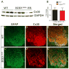
Cx43 levels are significantly increased in the spinal cord of SOD1G93A mice. (A) The protein level of Cx43 and GAPDH from lumbar spinal cord of control littermates at 60 days, 90 days and 120 days was examined. (B) Cx43 levels were elevated in the lumbar cord of SOD1G93A mice compared to control littler mate. (C) Cx43 expression was normalized to GAPDH and a significant increase was noted in SOD1G93A mice at endstage compared to control mice, with modest increases at 60 d and 90d. Data is presented as ±SEM with n =5 mice per group per time point. **p < 0.01 by ANOVA with Bonferroni post-hoc analysis. (D) Cx43 expression was also evaluated in the cervical, thoracic, and lumbar spinal cord segments of SOD1G93A and littermate control mice. A 2–3 fold increase was observed in all the segments at end stage of the SOD1G93A mice compared to the control mice sacrificed at similar age. Data is represented as ±SEM with n =5 mice per condition, per genotype and p < 0.05 by ANOVA with Bonferroni analysis. Localization of Cx43 in lumbar gray matter of WT control (F) and SOD1G93A mice (F′) was observed in astrocytes labeled intensely with GFAP in SOD1G93A mice (E′) compared to control mice (E) as shown in the merged images (G, G′). Scale bar =20μm.
FIGURE 2
FIGURE 2
Focal loss of Cx30 in the ventral gray matter of SOD1G93A mice. (A–B) Protein expression of Cx30 was examined in the end-stage tissue from lumbar spinal cord of SOD1G93A mice and WT control littermate at the same time point. Cx30 was normalized to GAPDH and no significant changes were observed between WT and SOD1G93A mice (n =3–5 mice/group). (C–E) Immunohistochemical analysis shows increased astrogliosis in SOD1G93A ventral gray matter (C′) compared to control gray matter (C). Interestingly, focal loss of Cx30 was noted in the lumbar gray matter of SOD1G93A mice (D′, E′) at endstage compared to uniform Cx30 expression in the gray matter of control mice (D, E). n =5 mice/group, scale bar= 100μm.
FIGURE 3
FIGURE 3
Human post-mortem tissues from ALS patients have elevated Cx43 expression. (A–C) NanoString analysis of motor cortex, cervical and lumbar spinal cord from control and sporadic ALS patients show increased Cx43 gene expression in ALS patients. Data is presented as ±SEM with n =5 patients per group, **p < 0.01 by t-test. The protein level of Cx43 was significantly greater in motor cortex (A′), cervical spinal cord (B′), and lumbar spinal cord (C′) of sporadic ALS patients compared to control patients. (D) The protein level of Cx43 that was normalized to GAPDH shows a ~2.5-fold increase in motor cortex of ALS patients, a 2-fold increase in cervical spinal cord and a ~8 fold increase in the lumbar spinal cord compared to control patients. Data is presented as ±SEM with n =5 patients per group per tissue. *p < 0.05, **p < 0.01, ***p < 0.001 by ANOVA with Bonferroni post-hoc analysis. (E) Cx43 increase was localized prominently to the gray matter of ALS cervical cord (right) compared to control cervical cord (left). Scale bar =80μm.
FIGURE 4
FIGURE 4
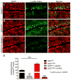
Cx43 expression is inherently elevated in mouse SOD1G93A astrocytes and ALS human iPSC-derived astrocytes. (A) Using qPCR analysis, a significant increase in Cx43 and GFAP levels was detected in SOD1G93A astrocytes compared to SOD1WT astrocytes, which was normalized with 18s RNA. (B) Similarly, increased protein levels of Cx43 and GFAP were detected in SOD1G93A astrocytes compared with SOD1WT astrocytes in vitro, which was normalized to the loading control actin. Data is presented as ±SEM, n =3 wells per condition. *p < 0.05, **p < 0.01 by ANOVA with Bonferroni analysis. (C) Human iPS cells from control and ALS patients were differentiated into astrocytes and stained with the astrocyte marker S100β along with Cx43. Scale bar =100μm (D) The iPSC-derived astrocytes from control (n =3) and ALS patients (n =6) were probed for Cx43 levels and was normalized to the loading control GAPDH. Data is presented as ±SEM for control lines and each of the ALS mutations are plotted individually showing higher Cx43 levels in SOD1A4V, C9ORF72 and sporadic patient lines **p < 0.01.
FIGURE 5
FIGURE 5
Increased Cx43 expression in SOD1G93A astrocytes mediates increased calcium responses through mechanical and ATP stimulation. (A) Mechanical stimulation of a single astrocyte with a glass micropipette at point of stimulation (black arrow, E1) generated calcium waves. (B) Quantification of calcium response in cells loaded with Fura-2 at 340 nm and 380 nm depicted increased calcium response of SOD1G93A astrocytes (red bar) compared with SOD1WT astrocytes (black bar) from point of stimulation E1 and different points away from point of stimulation. Data is presented as ±SEM with n =9–10 coverslips per condition and three independent experiments were conducted. *p < 0.05, **p < 0.01 between SOD1WT and SOD1G93A astrocytes by ANOVA with Bonferroni analysis. (C) Stimulation of astrocytes with 10 μm ATP for 30 sec results in elevated intracellular calcium levels, which returns back to baseline (after 45 secs). (D) Significantly higher intracellular calcium levels were observed in SOD1G93A astrocytes in response to ATP. This response was inhibited when the cells were incubated with the Cx43 blocker GAP26 in SOD1WT and SOD1G93A astrocytes. Data is presented as ±SEM with n =8–10 coverslips per condition and three independent experiments were conducted. *p < 0.05 between SOD1WT and SOD1G93A astrocytes and ^^^p < 0.001 between SOD1WT/SOD1G93A astrocytes and SOD1WT/SOD1G93A +peptide blocker by ANOVA with Bonferroni analysis.
FIGURE 6
FIGURE 6
Increased Cx43-mediated gap junction coupling occurs in SOD1G93A astrocytes. Scrape-loading assay was conducted to assess gap junction coupling using Lucifer yellow (LY) dye in SOD1WT astrocytes labeled with GFAP. Increased LY dye spread from the point of scrape was observed in SOD1G93A astrocytes (B) when compared with SOD1WT (A). (C) The spreading of LY dye was noticeably decreased when the cells were exposed to GAP26. (D) An appreciable increase in LY dye spreading and hence gap junctional coupling was observed in SOD1G93A astrocytes compared to control astrocytes. When SOD1WT and SOD1G93A astrocytes were treated with GAP26, LY dye spreading was significantly reduced with even further decline of signal in SOD1G93A astrocytes. Data is presented as ± SEM with n =4–5 coverslips per condition and three independent experiments were conducted. ***p < 0.001 between SOD1WT and SOD1G93A astrocytes, ^^p < 0.01 between SOD1WT and SOD1WT +GAP26, ^^^p < 0.01 between SOD1G93A and SOD1G93A +GAP26 by ANOVA followed by Bonferroni test. Scale bar =100 μm.
FIGURE 7
FIGURE 7
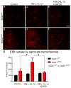
SOD1G93A astrocytes have increased hemichannel activity, which is heightened when stimulated with TNF-α and IL-1β. (A–B) Ethidium bromide (EtBr) uptake was conducted to measure the amount of hemichannel activity in SOD1WT and SOD1G93A astrocytes. (C–D) Astrocytes were stimulated with a cytokine mix of 10 μM of TNF-α and 10 μM of IL-1β for 24 hours and the hemichannel uptake was then measured the next day. (E, F) Astrocytes from SOD1WT and SOD1G93A mice were co-incubated with the cytokines and GAP26. (G) A significant increase was observed in EtBr uptake in SOD1G93A compared to SOD1WT astrocytes and this effect was further accentuated in SOD1G93A astrocytes when treated with cytokines. Notably, the inclusion of Cx43 blocker GAP26 diminishes this response back to baseline in SOD1G93A astrocytes. Data is represented as ±SEM with n =4–5 coverslips per condition and three independent experiments were conducted and analyzed using ANOVA. *p < 0.05, ***p < 0.001, SOD1WT compared to SOD1G93A astrocytes, #p < 0.05 SOD1G93A compared SOD1G93A +TNF-α +IL-1β, $p < 0.05 SOD1G93A +TNF-α +IL-1β compared to SOD1G93A +TNF-α +IL-1β+ GAP26. Scale bar =100 μm.
FIGURE 8
FIGURE 8
Blocking Cx43 in astrocyte-motor neuron co-culture is neuroprotective. (A) Motor neurons were plated on top of astrocytes from SOD1WT (A, A′) and SOD1G93A mice (B, B′) and SOD1G93A astrocytes treated with Cx43 hemichannel blocker GAP26 (C, C′) or with Cx43 blocker GAP19 (D, D′). Compared to motor neurons plated on SOD1WT astrocytes, motor neurons plated on top of SOD1G93A astrocytes degenerated rapidly (B, B′, E). However, when SOD1G93A astrocytes were treated with pan Cx43 blocker GAP26 (C, C′) or with Cx43 hemichannel blocker GAP19 (D, D′), significantly more motor neurons survived over time compared to untreated SOD1G93A astrocytes (E). Data is represented as ±SEM with n =4 wells per condition, per genotype and analyzed using ANOVA followed by Bonferroni post-hoc test. ^^^p < 0.01 between SOD1WT and SOD1G93A astrocytes, *p < 0.05, **p < 0.01 with ANOVA between SOD1G93A astrocytes and SOD1G93A astrocytes treated with GAP19 or GAP26. Scale bar =1000 μm.

References

    1. Abudara V, Bechberger J, Freitas-Andrade M, De Bock M, Wang N, Bultynck G, Naus CC, Leybaert L, Giaume C. The connexin43 mimetic peptide Gap19 inhibits hemichannels without altering gap junctional communication in astrocytes. Front Cell Neurosci. 2014;8:306. - PMC - PubMed
    1. Abudara V, Roux L, Dallerac G, Matias I, Dulong J, Mothet JP, Rouach N, Giaume C. Activated microglia impairs neuroglial interaction by opening Cx43 hemichannels in hippocampal astrocytes. Glia. 2015;63:795–811. - PubMed
    1. Avendano BC, Montero TD, Chavez CE, von Bernhardi R, Orellana JA. Prenatal exposure to inflammatory conditions increases Cx43 and Panx1 unopposed channel opening and activation of astrocytes in the offspring effect on neuronal survival. Glia. 2015;63:2058–2072. - PubMed
    1. Bennett MV, Contreras JE, Bukauskas FF, Saez JC. New roles for astrocytes: Gap junction hemichannels have something to communicate. Trends Neurosci. 2003;26:610–617. - PMC - PubMed
    1. Bruijn LI, Becher MW, Lee MK, Anderson KL, Jenkins NA, Copeland NG, Sisodia SS, Rothstein JD, Borchelt DR, Price DL, et al. ALS-linked SOD1 mutant G85R mediates damage to astrocytes and promotes rapidly progressive disease with SOD1-containing inclusions. Neuron. 1997;18:327–338. - PubMed

Publication types

MeSH terms