Review
. 2016 Apr:263 Suppl 1:S54-64.
doi: 10.1007/s00415-015-7903-4. Epub 2016 Apr 15.

Vestibular compensation: the neuro-otologist's best friend

Affiliations
Review

Vestibular compensation: the neuro-otologist's best friend

Michel Lacour et al. J Neurol. 2016 Apr.

Abstract

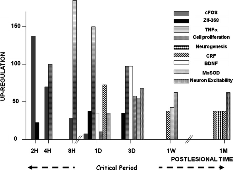
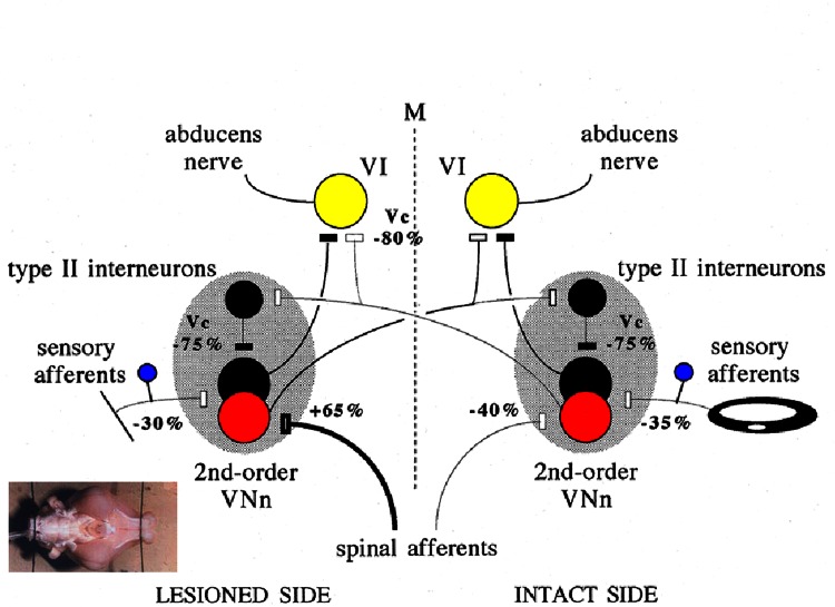
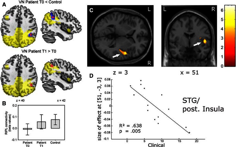
Why vestibular compensation (VC) after an acute unilateral vestibular loss is the neuro-otologist's best friend is the question at the heart of this paper. The different plasticity mechanisms underlying VC are first reviewed, and the authors present thereafter the dual concept of vestibulo-centric versus distributed learning processes to explain the compensation of deficits resulting from the static versus dynamic vestibular imbalance. The main challenges for the plastic events occurring in the vestibular nuclei (VN) during a post-lesion critical period are neural protection, structural reorganization and rebalance of VN activity on both sides. Data from animal models show that modulation of the ipsilesional VN activity by the contralateral drive substitutes for the normal push-pull mechanism. On the other hand, sensory and behavioural substitutions are the main mechanisms implicated in the recovery of the dynamic functions. These newly elaborated sensorimotor reorganizations are vicarious idiosyncratic strategies implicating the VN and multisensory brain regions. Imaging studies in unilateral vestibular loss patients show the implication of a large neuronal network (VN, commissural pathways, vestibulo-cerebellum, thalamus, temporoparietal cortex, hippocampus, somatosensory and visual cortical areas). Changes in gray matter volume in these multisensory brain regions are structural changes supporting the sensory substitution mechanisms of VC. Finally, the authors summarize the two ways to improve VC in humans (neuropharmacology and vestibular rehabilitation therapy), and they conclude that VC would follow a "top-down" strategy in patients with acute vestibular lesions. Future challenges to understand VC are proposed.

Keywords: Animal models; Dynamic deficits recovery; Human brain imaging; Static deficits recovery; Unilateral vestibular loss; Vestibular compensation.


Figures

Fig. 1
Fig. 1
Vestibular syndrome and recovery mechanisms in the compensation of acute unilateral vestibular loss. a The static ocular motor, postural and perceptive deficits are completely compensated by a rebalance of activity within the vestibular nuclei (VN) complexes on both sides. The orchestration of various neurobiological responses (or melodies) to VN deafferentation, depending on vestibular aetiology, is responsible for this re-balanced spontaneous firing between the VN on both sides. In contrast, the dynamic deficits (impaired vestibulo-ocular reflex and balance control in challenging conditions) are incompletely compensated. The whole brain functionally reorganizes and expresses new strategies (orchestration of behavioural responses or melodies) depending on the patients themselves. Neuropharmacology as well as vestibular rehabilitation therapy can alter the recovery of both the static and dynamic functions. b Main concepts explaining vestibular compensation: restoration, adaptation (sensory and behavioural substitutions) and habituation
Fig. 2
Fig. 2
Molecular and cellular mechanisms involved in vestibular compensation. The figure illustrates the plastic events occurring in the ipsilesional vestibular nuclei (VN) complex after acute unilateral vestibular loss. Up-regulation of the immediate early genes Fos and Zif-268 is found in the very first hours and days following the deafferentation, which induces a cascade of plastic events. Neurotrophic (BDNF) and neuroprotective (MnSOD) factors, markers of inflammation (TNFα), markers of the stress axis activation (corticotrophin-releasing factor: CRF) are up-regulated. Cell proliferation is seen very early (glial reaction, astrogenesis and neurogenesis) and is followed later on by cell differentiation leading particularly to newborn GABAergic neurons. Changes in intrinsic excitability of the VN neurons are observed also during the first post-lesional month, which could constitute an opportunity window, that is, a critical period for structural and functional reorganization in the ipsilesional VN complex. Up-regulations after vestibular lesion are expressed in percent of the basal level recorded in intact controls. Adapted from [, , –28, 80]
Fig. 3
Fig. 3
Plastic changes recorded in vitro that may underlie vestibular compensation in vivo. The figure (issued from [47]) shows the putative changes of synaptic efficacy, which have taken place in the vestibular-related pathways of compensated guinea pigs, 1 week after a unilateral labyrinthectomy, as they were recorded in an in vitro whole brain preparation of guinea pig. The percentages of modification of the response to single-shock stimulation of the various afferent inputs of MVNn and abducens motoneurons are shown in each case, while the thickness of the pathways has been reduced or increased accordingly. Vc indicates that the modified response was induced by stimulation of the contralateral vestibular nerve. The excitatory neurons and synapses are shown in white, whereas the inhibitory neurons and synapses are drawn in black. The absence of signs inside the neurons indicates balanced levels of spontaneous activity between both sides of the brain in these compensated guinea pigs. M median line of the brain, VI abducens motoneurons
Fig. 4
Fig. 4
Resting-state activity and voxel-based morphometry changes in patients with unilateral vestibular loss. a, b Resting-state activity changes (independent component analysis, component 50, beta values, with standard deviation, in arbitrary units) in right intraparietal sulcus (RIPL) contrasting 20 vestibular neuritis patients at acute unilateral vestibular failure (T0) and after 3 months (T1) with control subjects. Red area decreased circumscribed resting-state activity in patients (red p < 0.05, corrected; blue p < 0.005, uncorrected) which is partially reversed over time. Yellow component mask generated across both groups (patients and controls). Modified after [67]. c, d Voxel-based morphometry in patients with unilateral vestibulopathy due to surgical resection of acoustic neuroma: Significant correlation of gray matter volume (GMV) increases (ordinates) in the superior temporal gyrus (STG)/posterior insula with the clinical vestibular score (CVS, abscissae). CVS, a score reflecting vestibular impairment on clinical examination, is shown on representative axial and coronal slices of the standard MNI template and indicated by arrows (x-, z-coordinates below the slices). T-scores are indicated by the coloured map. The linear regression reflects that the increase in GMV is strongest with least clinical impairment. Modified after [70]

References

    1. Lacour M, Borel L. Vestibular control of posture and gait. Arch Ital Biol. 1993;131:81–104. - PubMed
    1. Wilson VJ, Melvill Jones GM. Mammalian vestibular physiology. New York, London: Plenum Press; 1979.
    1. Ferre ER, Longo MR, Fiori M, Haggard P. Vestibular modulation of spatial perception. Front Hum Neurosci. 2013;7:660. doi: 10.3389/fnhum.2013.00660. - DOI - PMC - PubMed
    1. Angelaki DE, Klier EM, Snyder LH. A vestibular sensation: probabilistic approaches to spatial perception. Neuron. 2009;64:448–461. doi: 10.1016/j.neuron.2009.11.010. - DOI - PMC - PubMed
    1. Lopez C, Lenngenhager B, Blank O. How vestibular stimulation interacts with illusory hand ownership. Conscious Cogn. 2010;19:33–47. doi: 10.1016/j.concog.2009.12.003. - DOI - PubMed