. 2016 Jun 15;11(2):029806.
doi: 10.1116/1.4944586.

Blood compatibility assessment of polymers used in drug eluting stent coatings

Affiliations

Blood compatibility assessment of polymers used in drug eluting stent coatings

Luisa Mayorga Szott et al. Biointerphases. .

Abstract

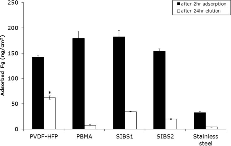
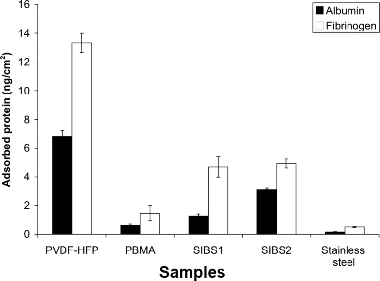
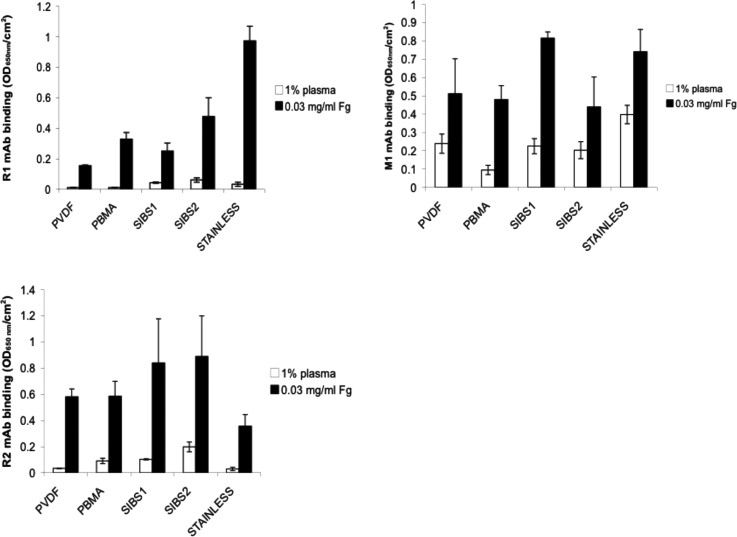
Differences in thrombosis rates have been observed clinically between different drug eluting stents. Such differences have been attributed to numerous factors, including stent design, injury created by the catheter delivery system, coating application technologies, and the degree of thrombogenicity of the polymer. The relative contributions of these factors are generally unknown. This work focuses on understanding the thrombogenicity of the polymer by examining mechanistic interactions with proteins, human platelets, and human monocytes of a number of polymers used in drug eluting stent coatings, in vitro. The importance for blood interactions of adsorbed albumin and the retention of albumin was suggested by the data. Microscopic imaging and immunostaining enhanced the interpretation of results from the lactate dehydrogenase cell counting assay and provided insight into platelet interactions, total quantification, and morphometry. In particular, highly spread platelets may be surface-passivating, possibly inhibiting ongoing thrombotic events. In many of the assays used here, poly(vinylidene fluoride-co-hexafluoropropylene) (PVDF-HFP) showed a differentiated protein deposition pattern that may contribute to the explanation of the consistently thromboresistant blood-materials interaction for fluororpolymers cited in literature. These results are supportive of one of several possible factors contributing to the good thromboresistant clinical safety performance of PVDF-HFP coated drug eluting stents.


Figures

F<sc>ig</sc>. 1.
Fig. 1.
Polymer structures for (a) PVDF-HFP, (b) poly(n-butyl methacrylate) (PBMA), and (c) polystyrene-b-polyisobutylene-b-polystyrene (SIBS1 and SIBS2).
F<sc>ig</sc>. 2.
Fig. 2.
Two-hour albumin adsorption from a pure Alb solution (0.3 mg/ml) in CPBSzI (black) and the retained Alb on the surfaces after a 24-h elution with 2% SDS (white). Data are expressed as mean ± SEM (n = 4). Single asterisks denote statistically significant differences in the amount of adsorbed Alb on to PVDF-HFP as compared to all other materials (α = 0.05). Double asterisks denote a significantly higher amount of retained Alb on PVDF-HFP as compared to all other materials studied (α = 0.05).
F<sc>ig</sc>. 3.
Fig. 3.
Two-hour fibrinogen adsorption from a pure Fg solution (0.03 mg/ml) in CPBSzI (black) and the retained Fg on the surfaces after a 24-hour elution with 2% SDS (white). Data are expressed as mean ± SEM (n = 4). The asterisk denotes the significantly greater amount of retained Fg on PVDF-HFP as compared to all other materials tested (α = 0.05).
F<sc>ig</sc>. 4.
Fig. 4.
Competitive protein adsorption from binary solutions (0.3 mg/ml Alb and 0.03 mg/ml Fg). Two hour protein adsorption data are represented by black bars or red bars, while 24 h elution data are represented by the white bars or pink bars. Data are presented as mean ± SEM (n = 4). There is a statistically significant higher amount of retained Alb on PVDF-HFP as compared to all other materials. There is a statistically significant higher of adsorbed Fg on SIBS-1 and SIBS-2 as compared to PVDF-HFP. There is a statistically significant higher amount of retained Fg on PVDF-HFP as compared to all other materials tested (α = 0.05 for all).
F<sc>ig</sc>. 5.
Fig. 5.
Two hour protein adsorption from 1% citrated normal human plasma. Alb adsorption is shown in black and Fg adsorption is shown in white. Data are displayed as mean ± SEM (n = 4). The single asterisk denotes statistically significant differences in Alb adsorption as compared to PVDF-HFP. Double asterisks denote statistically significant Fg adsorption as compared to PVDF-HFP (α = 0.05 for both).
F<sc>ig</sc>. 6.
Fig. 6.
Amount of retained protein from 1% citrated human plasma on the surface of the various materials after a 24-h elution with 2% SDS. Adsorbed Alb is shown in black and Fg is shown in white. Data are displayed as mean ± SEM (n = 4). Statistically significant differences in the amount of retained Alb and Fg, respectively, on PVDF-HFP as compared to all the other materials tested were noted (α = 0.05 for both).
F<sc>ig</sc>. 7.
Fig. 7.
Monoclonal antibody binding to samples preadsorbed with 1% pooled normal human citrated plasma (white bars) or 0.03 mg/ml Fg (black bars). The three antibodies used were selected for their specificity to three platelet binding sites on fibrinogen. R1 (a) and R2 (b) bind to RGD-containing regions on the alpha chain, while M1 (c) binds to the platelet binding dodecapeptide region on the gamma chain. Nonspecific binding was found to be minimal (data not shown). Data are displayed as mean ± SEM (n = 3).
F<sc>ig</sc>. 8.
Fig. 8.
Platelet adhesion to samples preadsorbed with 1% normal human citrated plasma measured using LDH assay. Data are displayed as mean ± SEM (n = 4).
F<sc>ig</sc>. 9.
Fig. 9.
Platelet adhesion on samples preadsorbed with pure 0.03 mg/ml pure Fg solution measured using LDH assay. Data are displayed as mean ± SEM (n = 4).
F<sc>ig</sc>. 10.
Fig. 10.
Scanning electron micrographs (2000× magnification) of adherent platelets onto samples preadsorbed with 1% plasma prior to exposing to PRP for 2 h.
F<sc>ig</sc>. 11.
Fig. 11.
Optical micrographs (40× magnification) of immunostained adherent platelets on PVDF-HFP, PBMA, SIBS1, and SIBS2 on 1% plasma preadsorbed surfaces.
F<sc>ig</sc>. 12.
Fig. 12.
Monocyte adhesion on samples preadsorbed with 1% plasma measured using LDH assay. Data are displayed as mean ± SEM (n = 4). Asterisks denote statistical significance as compared to stainless steel (α = 0.10).
F<sc>ig</sc>. 13.
Fig. 13.
No correlation was found between number of adherent platelets and M1 monoclonal antibody binding to surfaces preadsorbed with (a) plasma or (b) fibrinogen. Data are displayed as mean ± SEM (n = 3).

References

    1. Luscher T. F., Steffel J., Eberli F. R., Joner M., Nakazawa G., Tanner F. C., and Virmani R., Circulation 115, 1051 (2007).10.1161/CIRCULATIONAHA.106.675934 - DOI - PubMed
    1. Inoue T. and Node K., Circ. J. 73, 615 (2009).10.1253/circj.CJ-09-0059 - DOI - PubMed
    1. Vroman L., Adams A. L., and Fischer G. C., Adv. Chem. 199, 265 (1982).10.1021/ba-1982-0199 - DOI
    1. Slack S. M. and Horbett T. A., Proteins at Interfaces II: Fundamentals and Applications, edited by Brash J. L. and Horbett T. A. ( American Chemical Society, Washington, D.C., 1995).
    1. Brash J. L., “ Role of plasma protein adsorption in the response of blood to foreign surfaces,” in Blood Compatible Materials and Devices: Perspectives Towards the 21st Century, edited by Sharma C. P. and Szycher M. ( Technomic, Lancaster, PA, 1991).

Publication types

Substances

LinkOut - more resources