Review
. 2016 Apr 15;30(8):892-908.
doi: 10.1101/gad.277681.116.

Distinctive properties of metastasis-initiating cells

Affiliations
Review

Distinctive properties of metastasis-initiating cells

Toni Celià-Terrassa et al. Genes Dev. .

Abstract

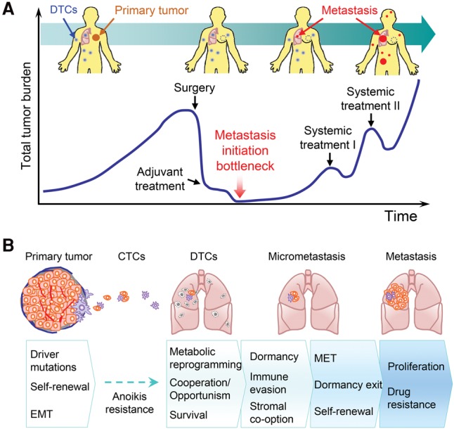
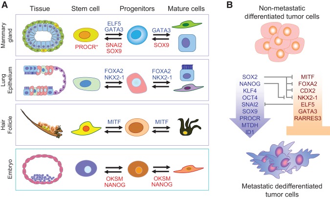
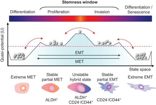
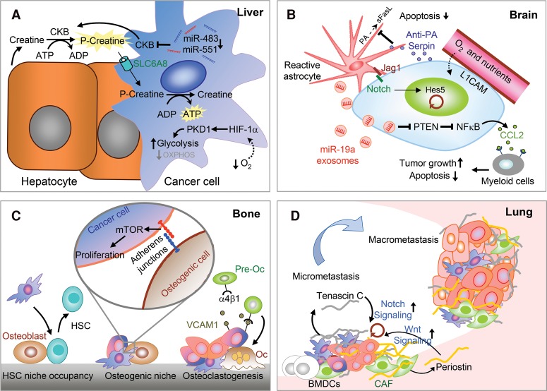
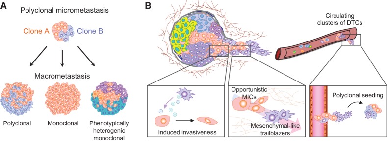
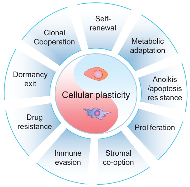
Primary tumors are known to constantly shed a large number of cancer cells into systemic dissemination, yet only a tiny fraction of these cells is capable of forming overt metastases. The tremendous rate of attrition during the process of metastasis implicates the existence of a rare and unique population of metastasis-initiating cells (MICs). MICs possess advantageous traits that may originate in the primary tumor but continue to evolve during dissemination and colonization, including cellular plasticity, metabolic reprogramming, the ability to enter and exit dormancy, resistance to apoptosis, immune evasion, and co-option of other tumor and stromal cells. Better understanding of the molecular and cellular hallmarks of MICs will facilitate the development and deployment of novel therapeutic strategies.

Keywords: cancer metastasis; epithelial–mesenchymal transition; metastasis-initiating cells; metastatic niche; plasticity.


Figures

Figure 1.
Figure 1.
Metastasis-initiating cells (MICs) in cancer progression and metastasis. (A) Schematic depiction of the typical course of metastatic progression of an early-stage cancer. In many clinical cases, tumor dissemination precedes diagnosis of the primary tumor. Surgical debulking and systemic adjuvant treatment eliminate most of tumor cells at the primary site and throughout the body. However, a small proportion of DTCs survives the systemic treatment. After a period of dormancy with no clinical sign of cancer, which could last for months to decades, clinically detectable metastases start to emerge. The subsequent lines of systemic treatment often only temporarily reduce the tumor burden before metastatic lesions develop resistance and eventually overwhelm the patients. The ability to initiate metastatic outgrowth is therefore a major bottleneck in cancer progression. (B) Representation of the sequence of events leading to metastasis initiation and acquisition of MIC properties. At the primary tumor site, a tiny fraction of long-term self-renewing tumor-initiating cells (TICs) may represent early MICs with driver mutations and high cellular plasticity. During dissemination, the large majority of DTCs dies, except those with strong anoikis resistance. Further attrition occurs after DTCs infiltrate distant organs, and MICs need to acquire a series of properties to become fully competent in seeding overt metastases.
Figure 2.
Figure 2.
Cell fate determinants in development and their influence on MICs. (A) Embryonic and adult epithelial cell lineage transcription factors tightly control self-renewal and lineage-specific differentiation of normal adult tissue stem cells and embryonic stem cells. (OKSM) The Yamanaka factors Oct4, Klf4, Sox2, and Myc. (B) The same transcription factors also influence metastatic behavior of cancer cells and the formation of MICs. Differentiation factors of normal tissues, such as MITF, GATA3, FOXA2, and others, act as tumor suppressors and metastasis inhibitors. On the other hand, dedifferentiation factors, such as the Yamanaka factors or tissue-specific stem cell factors, drive dedifferentiation, plasticity, and metastasis of MICs. Interestingly, these transcription factors constitute a complex network of reciprocal regulation. For example, SOX2 antagonizes multiple tissue-specific differentiation factors. Other factors, such as MTDH, exclusively support TIC and MIC activities and have no known function in normal tissue development.
Figure 3.
Figure 3.
Epithelial–mesenchymal plasticity and the “stemness window.” The schematic graph shows the putative multistable quasipotential landscape of epithelial–mesenchymal plasticity and the possible window of stemness in the transitional states. The vertical axis represents potential energy (U) differences between cell states, with higher potential corresponding to greater plasticity and stemness. The horizontal axis represents the state space gradient of epithelial and mesenchymal phenotypes. Gray balls represent populations of cells falling into different levels of potential energy, being more stable at lower levels. The red dashed line denotes a hypothetical threshold of minimal cell plasticity required to generate CSC activity. EMT and MET can lead MICs over the threshold of required potential energy for cellular plasticity. In fact, bidirectional transitions above this threshold would maintain MICs’ plasticity within the window of stemness in either a partial mesenchymal-like or epithelial-like state. Transitions between both partial states may experience a transitory high peak of potential energy and stemness, but this may represent a state of high instability. Extreme EMT or MET leads to a differentiated state impoverished of potential energy; therefore, cells falling at these states may completely lose plasticity and would not be capable of becoming MICs.
Figure 4.
Figure 4.
Cross-talk between MICs and stromal microenvironment and niches at different organs. (A) Metabolic adaptation in the liver. Low oxygen levels in the liver microenvironment force tumor cells to adapt via HIF-1α/PKD1 induction of glycolytic metabolism, thereby enabling metastatic colonization. MICs of colon cancer secrete CKB, which phosphorylates extracellular creatine produced by hepatocytes using extracellular ATP to generate phosphocreatine. Extracellular phosphocreatine is then imported into metastatic cancer cells by the transporter SLC6A8 to regenerate the ATP as a source of energy for survival and metastatic colonization. (B) Vascular niche and brain MIC–astrocyte cross-talk. The perivascular niche provides nutrients and oxygen to the infiltrating tumor cells, which secrete anti-PA serpins to protect MICs from astrocyte-derived death signals. Astrocytes also express Jagged1, which activates Notch signaling in MICs to promote self-renewal. Furthermore, astrocytes secrete miR-19a-containing exosomes, which suppress PTEN expression and activate CCL2-dependent recruitment of myeloid cells to promote tumor growth and survival in the brain. (C) In the bone, MICs compete with the hematopoietic stem cells (HSCs) for the HSC niche. Furthermore, osteogenic cells form heterotypic adherens junction with MICs and induce mTOR signaling to promote outgrowth. MICs also use secreted and membrane-bound VCAM1 to recruit preosteoclasts (pre-Oc) and activate their differentiation to mature osteoclasts (Oc), which in turn promote bone degradation and the formation of the “vicious cycle in bone metastasis.” (D) In the lung, bone marrow-derived cells (BMDCs) facilitate the formation of the premetastatic niche. In addition, the secretion of ECM proteins—tenascin C secreted by tumor cells and periostin (POSTN) secreted by stromal cells such as cancer-associated fibroblasts (CAFs)—further establishes the metastatic niche and supports MIC self-renewal by inducing Notch and Wnt signaling, respectively.
Figure 5.
Figure 5.
Clonal cooperation in metastasis. (A) Representation of different macrometastasis outputs from initial polyclonal dissemination and seeding. Polyclonal seeding and micrometastasis may develop polyclonal (left) or monoclonal (middle) macrometastasis depending on the clonal and tumor–stroma interaction dynamics in the target organ. (Right) In addition, metastasis heterogeneity can result from the generation of multiple phenotypes from a single metastatic clone. (B, left) Mesenchymal-like secretory cells can induce invasive phenotypes in epithelial-like TICs through secreted factors such as SPARC, facilitating their escape from the primary site. (Middle) In addition, noninvasive TICs/MICs can opportunistically follow trailblazer invasive cells to escape from the primary site or extravasate and infiltrate a distant tissue. (Right) Polyclonal seeding of a distant organ as a result of clonal cooperation.
Figure 6.
Figure 6.
Cellular plasticity as the core characteristic of MICs. This diagram lists nine characteristics commonly observed in MICs. These abilities are often enabled by cancer cell plasticity, represented as the core of MIC properties. Not all of these properties may need to be acquired simultaneously by MICs to grow metastases. Instead, multiple different combinations may influence the emergence of MICs in the affected organs.

References

    1. Aceto N, Bardia A, Miyamoto DT, Donaldson MC, Wittner BS, Spencer JA, Yu M, Pely A, Engstrom A, Zhu H, et al. 2014. Circulating tumor cell clusters are oligoclonal precursors of breast cancer metastasis. Cell 158: 1110–1122. - PMC - PubMed
    1. Acharyya S, Oskarsson T, Vanharanta S, Malladi S, Kim J, Morris PG, Manova-Todorova K, Leversha M, Hogg N, Seshan VE, et al. 2012. A CXCL1 paracrine network links cancer chemoresistance and metastasis. Cell 150: 165–178. - PMC - PubMed
    1. Aguilar E, Marin de Mas I, Zodda E, Marin S, Morrish F, Selivanov V, Meca-Cortés O, Delowar H, Pons M, Izquierdo I, et al. 2016. Metabolic reprogramming and dependencies associated with epithelial cancer stem cells independent of the epithelial–mesenchymal transition program. Stem Cells 10.1002/stem.2286. - DOI - PMC - PubMed
    1. Barcellos-Hoff MH, Lyden D, Wang TC. 2013. The evolution of the cancer niche during multistage carcinogenesis. Nat Rev Cancer 13: 511–518. - PubMed
    1. Beck B, Lapouge G, Rorive S, Drogat B, Desaedelaere K, Delafaille S, Dubois C, Salmon I, Willekens K, Marine JC, et al. 2015. Different levels of Twist1 regulate skin tumor initiation, stemness, and progression. Cell Stem Cell 16: 67–79. - PubMed

Publication types