Review
. 2017 Apr:151:101-138.
doi: 10.1016/j.pneurobio.2016.04.001. Epub 2016 Apr 12.

Monoaminergic neuropathology in Alzheimer's disease

Affiliations
Review

Monoaminergic neuropathology in Alzheimer's disease

Goran Šimić et al. Prog Neurobiol. 2017 Apr.

Abstract

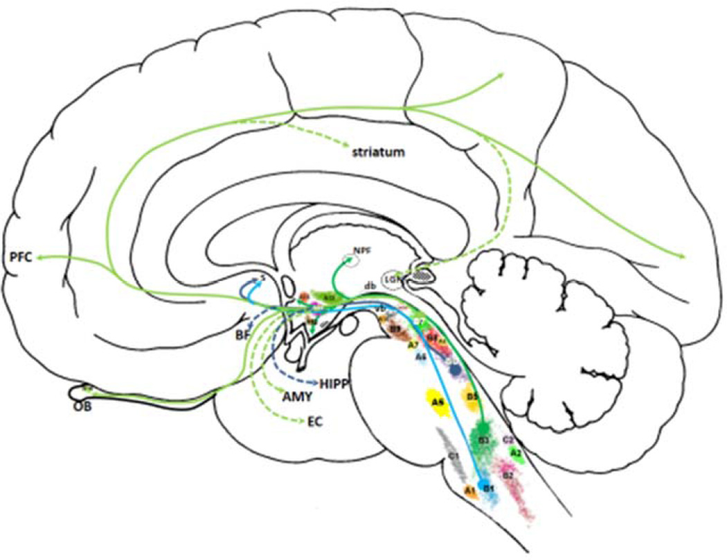
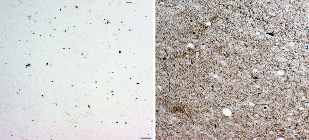
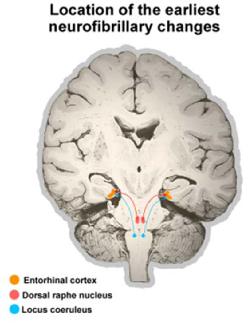
None of the proposed mechanisms of Alzheimer's disease (AD) fully explains the distribution patterns of the neuropathological changes at the cellular and regional levels, and their clinical correlates. One aspect of this problem lies in the complex genetic, epigenetic, and environmental landscape of AD: early-onset AD is often familial with autosomal dominant inheritance, while the vast majority of AD cases are late-onset, with the ε4 variant of the gene encoding apolipoprotein E (APOE) known to confer a 5-20 fold increased risk with partial penetrance. Mechanisms by which genetic variants and environmental factors influence the development of AD pathological changes, especially neurofibrillary degeneration, are not yet known. Here we review current knowledge of the involvement of the monoaminergic systems in AD. The changes in the serotonergic, noradrenergic, dopaminergic, histaminergic, and melatonergic systems in AD are briefly described. We also summarize the possibilities for monoamine-based treatment in AD. Besides neuropathologic AD criteria that include the noradrenergic locus coeruleus (LC), special emphasis is given to the serotonergic dorsal raphe nucleus (DRN). Both of these brainstem nuclei are among the first to be affected by tau protein abnormalities in the course of sporadic AD, causing behavioral and cognitive symptoms of variable severity. The possibility that most of the tangle-bearing neurons of the LC and DRN may release amyloid β as well as soluble monomeric or oligomeric tau protein trans-synaptically by their diffuse projections to the cerebral cortex emphasizes their selective vulnerability and warrants further investigations of the monoaminergic systems in AD.

Keywords: 5-hydroxytryptamine (serotonin); Alzheimer’s disease; Amyloid beta (Aβ) peptide; Blood-brain barrier; Cerebrospinal fluid; Epigenetics; Locus coeruleus; Metals; Monoamines; Neurofibrillary degeneration; Non-cognitive symptoms; Nucleus raphe dorsalis; Phosphorylation; Sleep-wake cycle; Tau protein.


Figures

Fig. 1
Fig. 1
Amyloid plaques, as revealed by Campbell-Switzer-Martin's method. The anterior part of the parahippocampal gyrus of an 84-year-old woman who died 3.5 years after the clinical diagnosis of AD was made. Scale bar = 1 mm.
Fig. 2
Fig. 2
Neurofibrillary changes in AD. Modified silver staining of the CA1 field from the body of the hippocampus of an 84-year-old woman, who died 3.5 years after clinical diagnosis of AD was made. NP = neuritic plaque. Numbers designate groups or "classes" of neurons with neurofibrillary changes, as defined and described in Braak et al., 1994b: 2 = early rod-like argyrophilic inclusions in the soma, 3 = typical developed NFT, which fills almost the whole cytoplasm and therefore acquires the shape of the neuron („flame-like“ appearance in the case of this pyramidal neuron), 4 = early extracellular NFT (such tangles are called „tombstone“ or „ghost“ tangles because the neurons have died and only the NFT remain), 5 = late extracellular tangle. Scale bar = 100 µm.
Figure 3
Figure 3
Vizualization of the hyperphosphorylated tau protein by using antibody AT8. A. The earliest detectable changes: hyperphosphorylated tau is localized in somatodendritic compartment of an isolated layer III pyramidal neuron in the transentorhinal cortex of a cognitively normal 59-year-old adult person. According to the criteria put forward in Braak et al. 1994b, such neurons belong to the group 1 neurons (they cannot be revealed by silver staining as they are bearing no tangle, just containing hyperphosphorylated tau). It is not known whether this change is reversible. B. Enlarged image from A. Note evenly distributed AT8-immunoreactive material in soma and all neuronal processes as well as grossly normal neuronal morphology. C. A group of temporal cortex pyramidal neurons in advanced stages of neurofibrillary degenerative changes in the brain of a 73-year old subject with a 7-year history of AD. A spectrum of conspicuous cytoskeletal alterations is visible in all five neurons (belonging to all groups/“classes“ neurons according to Braak et al., 1994, except group 5 end-stage neurons when their AT8 immunoreactivity is gone). All of these neurons also show argyrophilia, meaning that their neurofibrillary tangles can be revealed by using silver stainging methods. D. AT8 - immunoreactivity in 'granules' and tortuous apical and basal dendrites of granule cells of the hippocampal dentate gyrus of the same subject as in C. Perikarya of granule cells are rarely AT8-positive and do not usually contain typical NFT (even in cases with long-lasting history of AD), perhaps because this special neuronal type does not express MAPT mRNAs containing exon 10 (4R isoforms), supposedly conferring their resistance to neurofibrillary changes. Scale bars = 100 µm.
Fig. 4
Fig. 4
The Braak’s staging system (Braak and Braak, 1991). The topographic progression of AD classifies neurofibrillary degeneration in 6 stages, spreading from the transentorhinal region to the hippocampal formation (initial stages I and II, which clinically correlates with subjective or objective impairment of memory for recent events and mild spatial disorientation, but with preservation of general cognitive functioning without or with minimum impairment of activities of daily living), then to the temporal, frontal, and parietal neocortex (intermediate stages III and IV, which correlates with impaired recall, delayed word recall and word finding difficulties, disorientation in time and space, and impaired concentration, comprehension and conceptualization, among other symptoms of dementia), and finally to unimodal and primary sensory and motor areas of the neocortex (late stages V and VI, which roughly correlates with disturbances in object recognition, and other perceptual and motor skills). Braak staging system can be reduced to four with improved inter-rater reliability (Nagy et al., 1998): B0: no NFT, B1: Braak stages I/II, with NFT predominantly in entorhinal cortex and closely related areas, B2: stages III/IV, with NFTs more abundant in hippocampus and amygdala while extending slightly into the association cortex, and B3: stages V/VI, with NFT, neuropil threads and dystrophic neurites widely distributed throughout the neocortex and ultimately involving primary motor and sensory areas.
Figure 5
Figure 5
Schematic drawing of monoaminergic nuclei (except histaminergic and melatonergic cell groups). A1–A7 denote noradrenergic cell groups (A3 is missing in primates), A8–A16 dopaminergic cell groups (A11 is missing in humans, whereas retinal dopaminergic neurons are sometimes denoted as A17 group), B1–B9 serotonergic cell groups (B4 is missing in primates), whereas C1 and C2 denote adrenergic cell groups (C3 group is not present in humans). Histamine neurons in humans are located exclusively in tuberomammilary nucleus (stippled area caudal to A12). Melatonin neurons are located in the pineal gland (stippled area posterior to LGN). Emphasis is given on a rough sketch of two main ascending serotonergic systems: M-fibres with coarse varicosities take their origin from the nucleus raphe pontis (dorsalis, B6, purple lines) and nucleus raphe pallidus (B1, light blue lines) as well as from the nucleus raphe pontis medianus (B5) to a lesser extent (fibers from B5 not drawn) ascending through the tegmental area as the ventral bundle (vb), whereas fibres with small varicosities arise from NRD (light green lines, B7) and nucleus raphe magnus (dark green lines, B3) and collect in a dorsal bundle (db). Large serotonergic axons of the ventral bundle make synaptic contact with their targets, whereas fine axons with small varicosities release serotonin diffusely (volume transmission). AMY, amygdala; BF, basal forebrain; LGN, lateral geniculate nucleus; db, dorsal bundle of serotonergic fibers; EC, entorhinal cortex; HIPP, hippocampus; OB, olfactory bulb; NPF, nucleus parafascicularis; PFC, prefrontal cortex; S, septum; vb, ventral bundle of serotonergic fibers; see Table 1 for the designation of the serotonergic cells groups and their afferent and efferent connections. See text for detailed description.
Fig. 6
Fig. 6
Bielschowsky silver staining (left panel) and Gallyas silver iodide staining (right panel) of the supratrochlear part of the nucleus raphe dorsalis (NRD) of a 69-year-old woman with mild cognitive impairment (MCI), who had also documented several behavioral and psychological symptoms of dementia (BPSD). Although silver staining seems not to show many changes, on one of the adjacent sections from the same block of tissue more sensitive Gallyas silver iodide reveals a plethora of neurofibrillary changes, including NFT and degenerating neurites. Scale bars = 100 µm.
Figure 7
Figure 7
Schematic drawing of a speculative spreading of tau pathology from LC and DRN to the transentorhinal/entorhinal cortex.

References

    1. Adwan L, Zawia NH. Epigenetics: a novel therapeutic approach for the treatment of Alzheimer's disease. Pharmacol. Ther. 2013;139:41–50. (PubMed: 23562602) - PMC - PubMed
    1. Airaksinen MS, Paetau A, Paljarvi L, Reinikainen K, Riekkinen P, Suomalainen R, Panula P. Histamine neurons in human hypothalamus: anatomy in normal and Alzheimer diseased brains. Neuroscience. 1991;44:465–481. (PubMed: 1719449) - PubMed
    1. Albert MS, DeKosky ST, Dickson D, Dubois B, Feldman HH, Fox NC, Gamst A, Holtzman DM, Jagust WJ, Petersen RC, Snyder PJ, Carrillo MC, Thies B, Phelps CH. The diagnosis of mild cognitive impairment due to Alzheimer’s disease: recommendations from the National Institute on Aging – Alzheimer’s Association workgroups on diagnostic guidelines for Alzheimer’s disease. Alzheimers Dement. 2011;7:270–279. (PubMed: 21514249) - PMC - PubMed
    1. Alzheimer´s Association. Alzheimer’s Association Trial Match. [Accessed 17 Apr 2014]; http://www.alz.org/research/clinical_trials/find_clinical_trials_trialma....
    1. Ambree O, Richter H, Sachser N, Lewejohann L, Dere E, de Souza Silva MA, Herring A, Keyvani K, Paulus W, Schabitz WR. Levodopa ameliorates learning and memory deficits in a murine model of Alzheimer’s disease. Neurobiol. Aging. 2009;30:1192–1204. (PubMed: 18079024) - PubMed

Publication types