Review
. 2017 Jan;67(1):1-10.
doi: 10.1007/s12576-016-0450-7. Epub 2016 Apr 15.

The mammalian circadian clock and its entrainment by stress and exercise

Affiliations
Review

The mammalian circadian clock and its entrainment by stress and exercise

Yu Tahara et al. J Physiol Sci. 2017 Jan.

Abstract

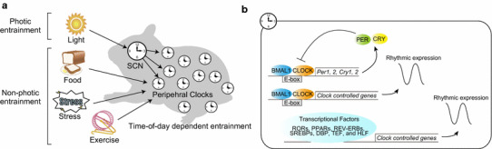
The mammalian circadian clock regulates day-night fluctuations in various physiological processes. The circadian clock consists of the central clock in the suprachiasmatic nucleus of the hypothalamus and peripheral clocks in peripheral tissues. External environmental cues, including light/dark cycles, food intake, stress, and exercise, provide important information for adjusting clock phases. This review focuses on stress and exercise as potent entrainment signals for both central and peripheral clocks, especially in regard to the timing of stimuli, types of stressors/exercises, and differences in the responses of rodents and humans. We suggest that the common signaling pathways of clock entrainment by stress and exercise involve sympathetic nervous activation and glucocorticoid release. Furthermore, we demonstrate that physiological responses to stress and exercise depend on time of day. Therefore, using exercise to maintain the circadian clock at an appropriate phase and amplitude might be effective for preventing obesity, diabetes, and cardiovascular disease.

Keywords: Liver; Mammalian circadian clock; Muscle; Oxidative stress.


Conflict of interest statement

The authors declare no competing financial interests.

Figures

Fig. 1
Fig. 1
Schematic diagram of the mammalian circadian clock. a External cues, such as light, food, stress, and exercise, entrain the central (suprachiasmatic nucleus; SCN) and peripheral (peripheral tissues) clocks. Light directly entrains the SCN, whereas other stimuli reset the peripheral clocks, and entrainment depends on the timing of stimulation. b The molecular clock is regulated by transcriptional feedback loops of core clock genes, and oscillations of clock-regulated genes in each tissue are controlled by various transcriptional factors, including CLOCK/BMAL1, RORs, PPARs, REV-ERBs, SREBPs, DBP, TEF, and HLF
Fig. 2
Fig. 2
Stress-induced phase shift of peripheral PER2::LUC rhythms. a Experimental schedule; 2-h restraint stress was performed for 3 days at Zeitgeber time (ZT)4–6 in PER2::LUC mice and, subsequently, the rhythm of in vivo bioluminescence was monitored. b Representative images of in vivo PER2::LUC bioluminescence in kidney (upper panels), liver, and submandibular gland (sub gla) tissues (lower panels). c Normalized PER2::LUC oscillations in control and stress groups show phase advancement in the stressed group. Values are expressed as mean ± SEM. The P values shown on the lower right side of the graphs indicate the results of two-way ANOVA (with Tukey post hoc test) between the control and stress groups. *P < 0.05, ***P < 0.001 (modified from [16])
Fig. 3
Fig. 3
Time-of-day dependence of circadian changes in response to restraint stress. a Phase-response curves of the response of peripheral clocks to 2-h restraint stress at Zeitgeber time (ZT)0–2, 4–6, 12–14, 16–18, and 20–22 (PER2::LUC rhythms). Increased and decreased phase shifts indicate phase-advance and -delay, respectively. Data for ZT25 were copied from ZT1. Graphs include all rhythmic and arrhythmic data. b Representative images of in vivo PER2::LUC bioluminescence (left) and normalized waveforms (right) after restraint stress at ZT0–2 for 3 days (modified from [16])
Fig. 4
Fig. 4
Decreased amplitude of PER2::LUC rhythms in peripheral tissues after daily caffeine injections. a Experimental schedule; mice were maintained under 12:12 h light–dark conditions and given intraperitoneal (IP) injections of saline (control; VEH) or 20 mg kg−1 caffeine (CAF) for 3 consecutive days at Zeitgeber time (ZT)1; in vivo monitoring was initiated at ZT9. b Normalized waveforms of PER2::LUC rhythms in saline- or caffeine-injected mice

References

    1. Bass J, Takahashi JS. Circadian integration of metabolism and energetics. Science. 2010;330(6009):1349–1354. doi: 10.1126/science.1195027. - DOI - PMC - PubMed
    1. Tahara Y, Shibata S. Chronobiology and nutrition. Neuroscience. 2013;253:78–88. doi: 10.1016/j.neuroscience.2013.08.049. - DOI - PubMed
    1. Moore RY, Eichler VB. Loss of a circadian adrenal corticosterone rhythm following suprachiasmatic lesions in the rat. Brain Res. 1972;42(1):201–206. doi: 10.1016/0006-8993(72)90054-6. - DOI - PubMed
    1. Tahara Y, Kuroda H, Saito K, Nakajima Y, Kubo Y, Ohnishi N, Seo Y, Otsuka M, Fuse Y, Ohura Y, Komatsu T, Moriya Y, Okada S, Furutani N, Hirao A, Horikawa K, Kudo T, Shibata S. In vivo monitoring of peripheral circadian clocks in the mouse. Curr Biol. 2012;22(11):1029–1034. doi: 10.1016/j.cub.2012.04.009. - DOI - PubMed
    1. Panda S, Antoch MP, Miller BH, Su AI, Schook AB, Straume M, Schultz PG, Kay SA, Takahashi JS, Hogenesch JB. Coordinated transcription of key pathways in the mouse by the circadian clock. Cell. 2002;109(3):307–320. doi: 10.1016/S0092-8674(02)00722-5. - DOI - PubMed