. 2016 May 19;44(9):3989-4004.
doi: 10.1093/nar/gkw271. Epub 2016 Apr 15.

The DBHS proteins SFPQ, NONO and PSPC1: a multipurpose molecular scaffold

Affiliations

The DBHS proteins SFPQ, NONO and PSPC1: a multipurpose molecular scaffold

Gavin J Knott et al. Nucleic Acids Res. .

Abstract

Nuclear proteins are often given a concise title that captures their function, such as 'transcription factor,' 'polymerase' or 'nuclear-receptor.' However, for members of the Drosophila behavior/human splicing (DBHS) protein family, no such clean-cut title exists. DBHS proteins are frequently identified engaging in almost every step of gene regulation, including but not limited to, transcriptional regulation, RNA processing and transport, and DNA repair. Herein, we present a coherent picture of DBHS proteins, integrating recent structural insights on dimerization, nucleic acid binding modalities and oligomerization propensity with biological function. The emerging paradigm describes a family of dynamic proteins mediating a wide range of protein-protein and protein-nucleic acid interactions, on the whole acting as a multipurpose molecular scaffold. Overall, significant steps toward appreciating the role of DBHS proteins have been made, but we are only beginning to understand the complexity and broader importance of this family in cellular biology.


Figures

Figure 1.
Figure 1.
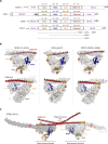
DBHS protein domain architecture and structure. (A) Schematic representation of DBHS protein domain architecture with the structurally characterized RNA recognition motifs (RRM) RRM1 (brown) and RRM2 (blue), NonA/ paraspeckle (NOPS) (orange) and coiled-coil (red) domains indicated. The uncharacterized DNA-binding domain (DBD) of SFPQ and other low complexity regions of each paralog are indicated in dashed boxes. The RGG motifs are represented in green within the SFPQ schematic. The corresponding amino acid boundaries for each protein are indicated above the schematic for NONO-1 (C. elegans), SFPQ, NONO and PSPC1 (H. sapiens). (B) X-ray crystal structures of NONO-1 (5CA5) (2), SFPQ (4WII) (12) and PSPC1/NONO (3SDE) (11). The first subunit of each dimer is illustrated as a domain colored cartoon and the second subunit as a molecular surface (gray). Directly below is an additional representation of each dimer from an identical perspective where the converse is shown; the first subunit of the dimer is illustrated as a surface (gray) and the second subunit of the dimer illustrated as a domain colored cartoon. (C) X-ray crystal structure of the SFPQ homodimer (4WIJ) (12) illustrating two SFPQ homodimers interacting via their coiled-coil oligomerization motif (highlighted by a red mark). The additional coiled-coil interaction sites within partnered chains are highlighted by a red mark. The dimerization domain and coiled-coil mediated oligomerization site are indicated. Prime (′) denotes the partner chain. Domains are colored consistently throughout.
Figure 2.
Figure 2.
DBHS protein-binding sites and post-translation modifications mapped to the X-ray crystal structure of SFPQ (4WIJ). The structure illustrates a putative SFPQ/NONO heterodimer (colored surface/black cartoon, respectively) with the remaining N- and C-terminal uncharacterized and low-complexity domains modeled as flexible chains at the corresponding termini of the structure (dashed lines). Interaction sites within the X-ray crystal structure are colored; dimerization interface (green), coiled-coil oligomerization motif (yellow), secondary oligomerization site (brown), putative RNA-binding surface of RRM1 (light blue), putative RNA-binding loop of RRM2 (dark blue). The structurally uncharacterized DNA-binding domain (12) of SFPQ is also illustrated (purple box). Mapped as colored circles to the SFPQ and NONO chains are reported sites of post-translational modification and corresponding amino acid number; phosphorylation (red), methylation (orange), citrullination (teal), SUMOylation (purple) and ADP-ribosylation (pale green). Methylation sites that are also subject to citrullination are indicated with an asterisk.
Figure 3.
Figure 3.
Simplified schematic representation of DBHS protein function. The DBHS proteins SFPQ (S) and NONO (N) are represented as simple red and green spheres respectively. (1) SFPQ and NONO can sequester transcription factors away from target promoters, (2) act as co-repressors at target promoters and (3) in complex with repressors stimulate epigenetic silencing. (4) Both SFPQ and NONO are associated with co-activation of transcription through (5) elongation up to termination. (6) SFPQ and NONO also remain associated with nascent mRNA to facilitate co-transcriptional processing, (7) mRNP export and (8) cytosolic trafficking. (9,10) By virtue of their involvement in paraspeckle formation and integrity, SFPQ and NONO can facilitate nuclear RNA retention. SFPQ, NONO and PSPC1 are also involved in double stranded break repair (11).
Figure 4.
Figure 4.
Context-dependent functions of mixed dimers and oligomers of DBHS protein with clinical significance. Broadly, DBHS proteins function in the clinical contexts of development, innate immunity and cancer. Corresponding examples are shown in colored boxes. The functional or mechanistic manifestation of that clinical context is indicated with a line. Note that paraspeckles may be involved in regulating DBHS protein partitioning for the other functions shown (indicated by dashed lines).

References

    1. Dong B., Horowitz D.S., Kobayashi R., Krainer A.R. Purification and cDNA cloning of HeLa cell p54nrb, a nuclear protein with two RNA recognition motifs and extensive homology to human splicing factor PSF and Drosophila NONA/BJ6. Nucleic Acids Res. 1993;21:4085–4092. - PMC - PubMed
    1. Knott G.J., Lee M., Passon D.M., Fox A.H., Bond C.S. Caenorhabditis elegans NONO-1: Insights into DBHS protein structure, architecture and function. Protein Sci. 2015;24:2033–2043. - PMC - PubMed
    1. Shav-Tal Y., Zipori D. PSF and p54(nrb)/NonO - multi-functional nuclear proteins. FEBS Lett. 2002;531:109–114. - PubMed
    1. Bond C.S., Fox A.H. Paraspeckles: nuclear bodies built on long noncoding RNA. J. Cell. Biol. 2009;186:637–644. - PMC - PubMed
    1. Fox A.H., Lamond A.I. Paraspeckles. Cold Spring Harbor Perspect. Biol. 2010;2 - PMC - PubMed

MeSH terms

LinkOut - more resources