. 2016 Jul 8;44(12):5702-16.
doi: 10.1093/nar/gkw275. Epub 2016 Apr 15.

Tetratricopeptide repeat factor XAB2 mediates the end resection step of homologous recombination

Affiliations

Tetratricopeptide repeat factor XAB2 mediates the end resection step of homologous recombination

David O Onyango et al. Nucleic Acids Res. .

Abstract

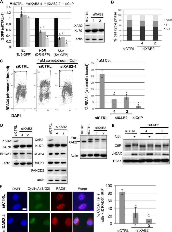
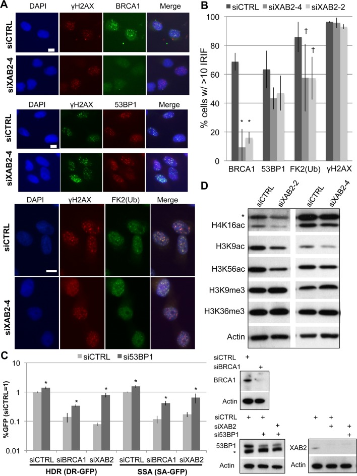
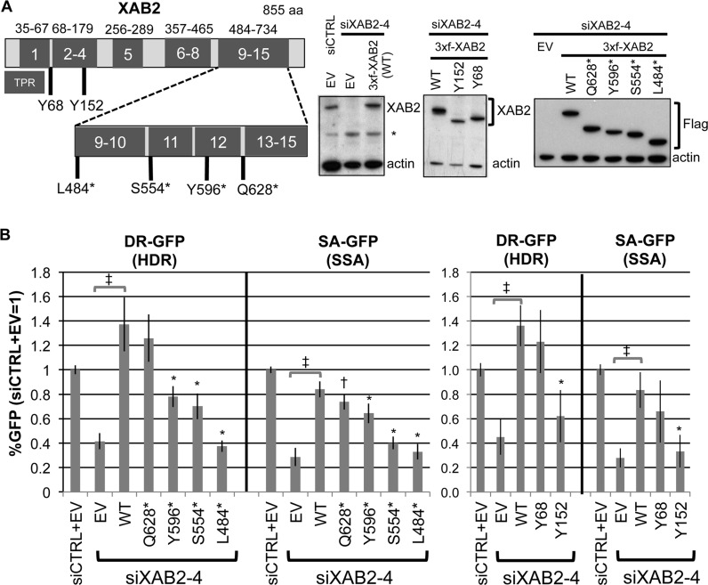
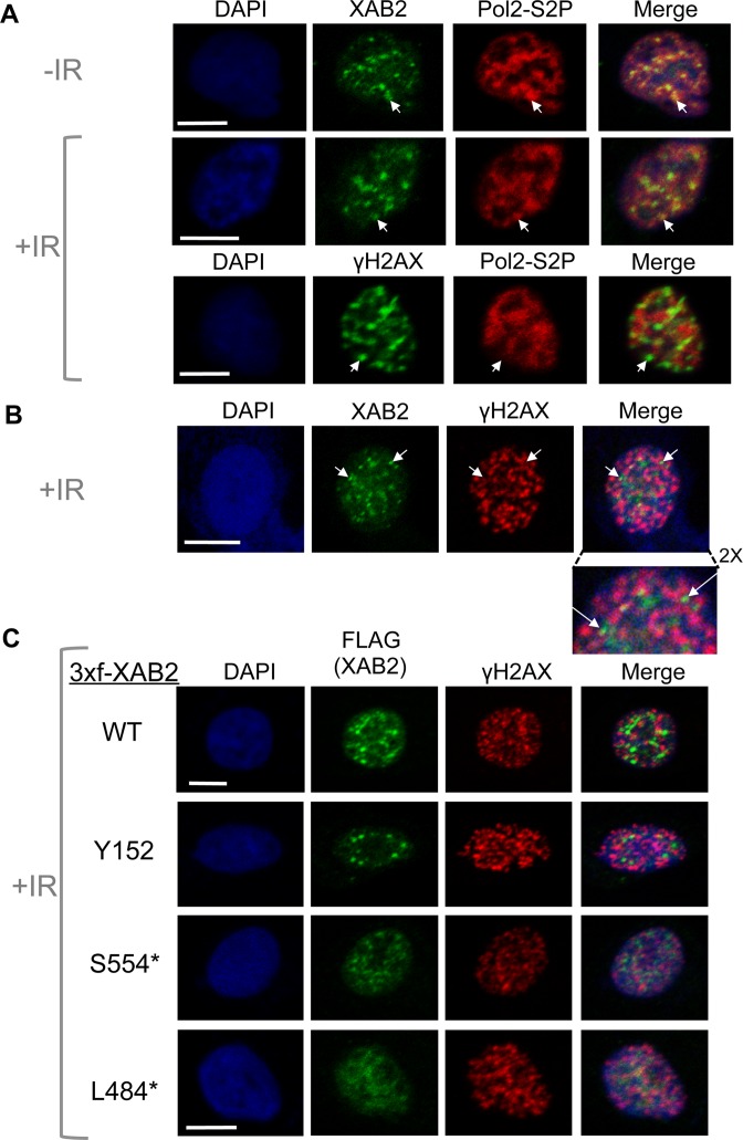
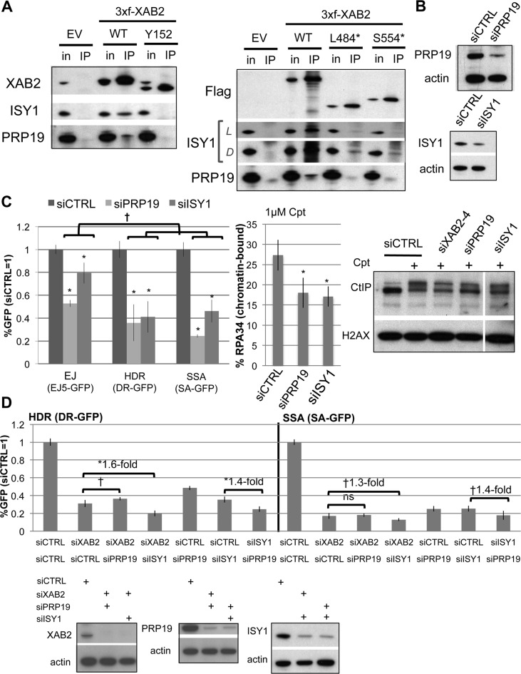
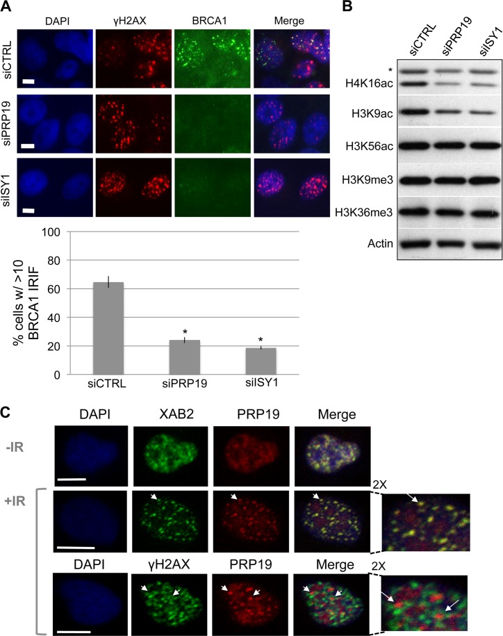
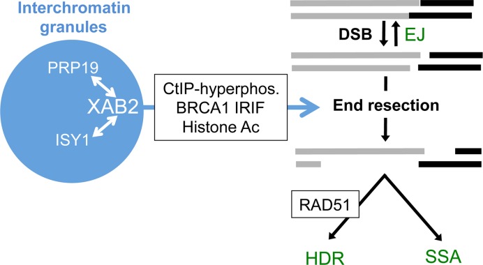
We examined the influence of the tetratricopeptide repeat factor XAB2 on chromosomal break repair, and found that XAB2 promotes end resection that generates the 3' ssDNA intermediate for homologous recombination (HR). Namely, XAB2 is important for chromosomal double-strand break (DSB) repair via two pathways of HR that require end resection as an intermediate step, end resection of camptothecin (Cpt)-induced DNA damage, and RAD51 recruitment to ionizing radiation induced foci (IRIF), which requires end resection. Furthermore, XAB2 mediates specific aspects of the DNA damage response associated with end resection proficiency: CtIP hyperphosphorylation induced by Cpt and BRCA1 IRIF. XAB2 also promotes histone acetylation events linked to HR proficiency. From truncation mutation analysis, the capacity for XAB2 to promote HR correlates with its ability to form a complex with ISY1 and PRP19, which show a similar influence as XAB2 on HR. This XAB2 complex localizes to punctate structures consistent with interchromatin granules that show a striking adjacent-localization to the DSB marker γH2AX. In summary, we suggest that the XAB2 complex mediates DNA damage response events important for the end resection step of HR, and speculate that its adjacent-localization relative to DSBs marked by γH2AX is important for this function.


Figures

Figure 1.
Figure 1.
XAB2 is important for HR DSB repair pathways that require end resection (HDR and SSA), Cpt-induced end resection, hyper-phosphorylation of CtIP, and RAD51 IRIF. (A) RNAi-depletion of XAB2 in U2OS cells causes a reduction in HDR and SSA, but not EJ. Shown are immunoblot signals for XAB2 and two loading controls (Ku70 and actin) after transfection with a non-targeting siRNA (siCTRL), two XAB2 siRNAs (siXAB2-2 and siXAB2-4), and a CtIP siRNA. Shown are the frequencies of GFP+ cells for the EJ5-GFP (EJ), DR-GFP (HDR), and SA-GFP (SSA) reporter lines transfected with an I-SceI expression vector after pre-treatment with the siRNAs shown. GFP+ frequencies are normalized to parallel siCTRL treated samples (= 1). Distinct from siCTRL, *P ≤ 0.0001, P = 0.013 (N = 6). (B) XAB2 depletion does not obviously affect cell cycle phase distribution. Shown are the percentage of cells in G1, S and G2/M based on propidium iodide/BrdU staining, after treatment with the siRNAs shown, and pulse BrdU labeling (N = 2). (C) XAB2 depletion causes a reduction of Cpt-induced end resection. Following siRNA treatment, cells were treated with Cpt, and mild detergent extracted prior to fixation and staining with RPA34 and DAPI. Shown are representative flow cytometry plots for cells treated with siCTRL and siXAB2-4, as well as the percentage of cells showing detergent resistant (i.e. chromatin bound) RPA34 staining. *Distinct from siCTRL, P < 0.0001, N ≥ 4. (D) XAB2 depletion does not have an obvious effect on the levels of several DDR proteins, except a modest decrease in BRCA1 for only siXAB2-2. Shown are immunoblot signals for several DDR factors for cells treated with the siRNAs shown. (E) XAB2 promotes CtIP hyper-phosphorylation induced by Cpt treatment. Following siRNA treatment, cells were treated with Cpt, and then treated with mild detergent prior to extraction of chromatin-bound proteins. Shown are CtIP, γH2AX and H2AX immunoblotting signals from representative samples. (F) XAB2 depleted cells show reduced RAD51 IRIF. For IRIF analysis, following siRNA treatment, cells were exposed to 10 Gy IR, and allowed to recover for 6 h prior to fixation for immunofluorescence analysis. Shown are representative images of RAD51 and Cyclin-A (S/G2 marker) staining of siCTRL and siXAB2-4 treated cells (scale bar = 10 μm), as well as the percentage of Cyclin-A+ cells showing >10 RAD51 IRIF for each siRNA treatment. *Distinct from siCTRL, P < 0.004 (N = 3).
Figure 2.
Figure 2.
XAB2 promotes BRCA1 IRIF and histone acetylation events associated with HR proficiency, and HR defects in siXAB2 treated cells can be suppressed by 53BP1 depletion. (A) XAB2 depleted cells show markedly reduced BRCA1 IRIF, a minor reduction in ubiquitin-chain IRIF, and no statistical decrease on 53BP1 or γH2AX IRIF. IRIF experiments were performed as in Figure 1F. Shown are representative images of BRCA1, 53BP1, ubiquitin-chain (FK2), and γH2AX staining of siCTRL and siXAB2-4 treated cells (scale bar = 10 μm). (B) Shown is the percentage cells showing >10 IRIF for each marker shown in (A), and for each siRNA treatment (N ≥ 3). Distinct from siCTRL: *P < 0.002 (N = 3), P < 0.04 (N = 5). (C) Treating cells with siRNAs targeting 53BP1 can suppress the HR defects of cells depleted of XAB2. DR-GFP and SA-GFP reporter cells were treated with several double siRNA combinations, maintaining the same total siRNA concentration as experiments in Figure 1A, prior to I-SceI expression, and analysis of GFP+ cells (siXAB2; siXAB2-4). The I-SceI transfection was 2 days after siRNA treatment for the DR-GFP experiments to allow for efficient RNAi depletion of 53BP1. Shown are the frequencies of GFP+ cells from these experiments, relative to siCTRL (= 1). *Distinct from the siRNA treatment without si53BP1, P ≤ 0.0007, (HDR N = 5; SSA N = 6). Immunoblots confirming depletion of the target protein by the respective siRNAs are also shown (*non-specific band). (D) XAB2 promotes histone acetylation levels. Following siRNA treatment, cells were treated with mild detergent prior to extraction of chromatin-bound proteins, as in Figure 1E. Shown are immunoblotting signals from representative samples for several histone modifications; *band detected by the H4K16Ac antibody that is non-specific, in that it migrates much slower than histone H4.
Figure 3.
Figure 3.
Defining regions of the XAB2 protein that are important to promote HR using truncation mutant analysis. (A) Expression of XAB2 mutant forms. Shown is a diagram of the XAB2 protein, with TPR motif regions outlined in dark grey, along with the positions of a series of truncation mutations (not to scale). All expression cassettes begin with an N-terminal 3xflag tag (3xf) and silent mutations to resist siXAB2-4. Mutants Y68 and Y152 represent a deletion (Δ) of 1–67 and 1–151, respectively, and the asterisks in the other mutants represent stop codons in place of the residue. Shown are immunoblot signals for XAB2 or Flag from cells transfected with various XAB2 expression vectors or EV following treatment with siXAB2-4 or siCTRL. (B) Distinct XAB2 truncation mutants show a varying capacity to promote HR: Y152 (Δ1–151) and L484* (Δ484-855) are deficient, S554* (Δ554–866) and Y596* (Δ596–855) show partial deficiency, whereas Y68 (Δ1–67) and Q628* (Δ628–855) are proficient. The DR-GFP (HDR) and SA-GFP (SSA) U2OS reporter cell lines were treated with siCTRL or siXAB2-4, and subsequently co-transfected with the I-SceI expression vector combined with the various XAB2 expression vectors shown in (A), or EV. Shown are the frequencies of GFP+ cells from these experiments, relative to parallel siCTRL/EV transfections (= 1). XAB2-WT distinct from EV: P≤0.0001. Mutants distinct from XAB2-WT: *P < 0.001, P = 0.02.
Figure 4.
Figure 4.
XAB2 shows punctate nuclear staining, which overlaps with a phosphorylated form of RNA polymerase II (Pol2-S2P), but shows a striking adjacent-localization to the DSB marker γH2AX (i.e. XAB2 and γH2AX often localize to adjacent sites). Cells were either treated with 10 Gy IR (+IR) or mock treated (-IR) and allowed to recover for 30 min prior to mild detergent extraction and fixation for immunofluorescence analysis using confocal microscopy. Scale bars = 10 μm. (A) Shown are immunofluorescence signals for representative cells: -IR treated and stained for Pol2-S2P and XAB2, +IR treated and stained for Pol2-S2P and XAB2, and +IR treated and stained for Pol2-S2P and γH2AX. Arrows in the top panels highlight examples of bright Pol2-S2P signal that co-localizes with punctate XAB2 staining, whereas those in the bottom panel highlight regions of low Pol2-S2P signal with bright γH2AX staining. (B) Shown are immunofluorescence signals for XAB2 and γH2AX of a representative cell, where arrows highlight two examples of punctate XAB2 signals that are adjacent to γH2AX stained regions. Also shown is a 2X magnification of the top of this cell. (C) Exogenously expressed XAB2 shows a similar staining pattern as the endogenous protein, whereas a C-terminal truncation mutant that is deficient in HR (L484*) shows more diffuse nuclear staining. Shown are immunofluorescence signals of Flag and γH2AX for representative cells treated with siXAB2-4, and then transfected with expression vectors for 3xf-XAB2 WT, Y152, S554* and L484*.
Figure 5.
Figure 5.
XAB2 forms a complex with PRP19 and ISY1, which also promote end resection. (A) XAB2-WT, but not HR-deficient truncation mutants, forms a complex with PRP19 and ISY1. Extracts were prepared from cells with transient expression of 3xf-XAB2 WT, Y152, L484* and S544*, which were treated with Cpt prior to harvesting. A fraction of each extract was used for input analysis (in), and the rest was used for a Flag immunoprecipitation (IP). Shown are immunoblot signals for XAB2, ISY1, PRP19, and Flag (the latter for the C-terminal truncation mutants). The PRP19 immunoblots also show a smaller non-specific band (i.e. also in the EV IP), which is likely due to signal from the Flag IP antibody. L = Light exposure, D = Dark exposure. (B) Depletion of ISY1 and PRP19 with siRNA. Shown are immunoblot signals for cells treated with siRNAs targeting ISY1 (siISY1) and PRP19 (siPRP19). (C) PRP19 and ISY1 affect HR similarly to XAB2. The influence of siISY1 and siPRP19 treatment on DSB reporter assays, end resection of Cpt-induced damage (chromatin-bound RPA34), and CtIP-hyperphosphorylation was performed as described in Figure 1E. Shown are repair values from the reporter assays (N ≥ 5), *distinct from siCTRL P < 0.0001, P<0.0001 except P = 0.04 for siPRP19 regarding HDR versus EJ. Shown is the percentage of RPA34+ cells after Cpt treatment for each siRNA treatment, *P < 0.0001, N = 9. Also shown are CtIP immmunoblot signals for cells treated with the siRNAs shown, that were subsequently Cpt treated (+), or untreated (–). (D) Double siRNA treatment combinations targeting XAB2 (siXAB2-4), ISY1, and PRP19, each cause a fold-decrease in HR that is less than the addition of the fold-decrease caused by the individual siRNA treatments (i.e. not additive). DSB reporter cell lines were treated with a series of double siRNA combinations, maintaining the same total siRNA concentration as experiments in Figure 1A, prior to I-SceI expression. Shown are the frequencies of GFP+ cells from these experiments, relative to siCTRL (= 1). Also shown is the fold-decrease in HR for double siRNA treatments relative to the respective individual siRNA treatment, which in each case are less than 2-fold. In contrast, each of the individual siRNA treatments cause a decrease in HR that is at least 2-fold. P = 0.015, *P < 0.0001 (N = 6). Also shown are immunoblots confirming depletion of the target proteins by the respective double siRNA treatments.
Figure 6.
Figure 6.
As with XAB2, ISY1 and PRP19 promote BRCA1 IRIF and histone acetylation. (A) PRP19 and ISY1 promote BRCA1 IRIF. Cells were treated with siRNAs targeting PRP19 and ISY1 as in Figure 5B, and subsequently BRCA1 IRIF were analyzed by immunofluorescence as in Figure 2A. Shown are representative cells stained for γH2AX and BRCA1, as well as the frequency of cells showing BRCA1 IRIF for cells treated with the respective siRNAs. *P < 0.0001 (N = 3). Scale bars = 10 μm. (B) PRP19 and ISY1 are important for H3K9Ac and H4K16Ac levels. Cells were treated with siRNAs targeting PRP19 and ISY1 as in (A), and modified histones were analyzed as in Figure 2D. Shown are representative immunoblot signals for several modified histones and actin from cells treated with the respective siRNAs. (C) PRP19 and XAB2 show substantial co-localization, and PRP19 shows adjacent-localization staining relative to γH2AX. Cells were treated as in Figure 4, and analyzed by immunostaining. Scale bars = 10 μm. Shown are immunofluorescence signals of representative cells for XAB2 and PRP19, and γH2AX and PRP19. Arrows highlight examples of punctate PRP19 signals that co-localize with XAB2 (middle panel), and are adjacent to γH2AX stained regions (bottom panel). Also shown are 2X magnification images from a top segment of the +IR treated cells.
Figure 7.
Figure 7.
A model for the influence of XAB2 and associated factors on the end resection step of HR. XAB2, along with PRP19 and ISY1, are depicted localizing to interchromatin granules that are apart from DSBs. XAB2 is depicted as promoting a set of DDR events linked to end resection of DSBs, which are important for both HDR (and RAD51 IRIF) and SSA, but are dispensable for EJ.

References

    1. Roy R., Chun J., Powell S.N. BRCA1 and BRCA2: different roles in a common pathway of genome protection. Nat. Rev. Cancer. 2012;12:68–78. - PMC - PubMed
    1. Kass E.M., Jasin M. Collaboration and competition between DNA double-strand break repair pathways. FEBS Lett. 2010;584:3703–3708. - PMC - PubMed
    1. San Filippo J., Sung P., Klein H. Mechanism of eukaryotic homologous recombination. Annu. Rev. Biochem. 2008;77:229–257. - PubMed
    1. Lin F.L., Sperle K., Sternberg N. Model for homologous recombination during transfer of DNA into mouse L cells: role for DNA ends in the recombination process. Mol. Cell. Biol. 1984;4:1020–1034. - PMC - PubMed
    1. Symington L.S., Gautier J. Double-strand break end resection and repair pathway choice. Annu. Rev. Genet. 2011;45:247–271. - PubMed

MeSH terms