. 2016 Jun;186(6):1547-58.
doi: 10.1016/j.ajpath.2016.02.004. Epub 2016 Apr 13.

Interferon-γ-Induced Unfolded Protein Response in Conjunctival Goblet Cells as a Cause of Mucin Deficiency in Sjögren Syndrome

Affiliations

Interferon-γ-Induced Unfolded Protein Response in Conjunctival Goblet Cells as a Cause of Mucin Deficiency in Sjögren Syndrome

Terry G Coursey et al. Am J Pathol. 2016 Jun.

Abstract

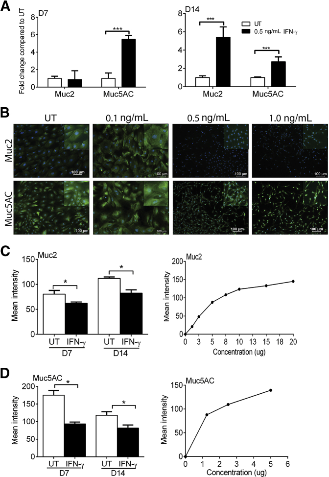
Goblet cells (GCs) are specialized secretory cells that produce mucins and a variety of other proteins. Significant conjunctival GC loss occurs in both experimental dry eye models and patients with keratoconjunctivitis sicca due to the induction of interferon (IFN)-γ. With the use of a primary murine culture model, we found that GCs are highly sensitive to IFN-γ with significantly reduced proliferation and altered structure with low concentrations. GC cultures treated with IFN-γ have increased gene expression of Muc2 and Muc5AC but do not express these mucin glycoproteins. We hypothesized that IFN-γ induces endoplasmic reticulum stress and the unfolded protein response (UPR) in GCs. Cultures treated with IFN-γ increased expression of UPR-associated genes and proteins. Increased GRP78 and sXBP1 expression was found in experimental dry eye and Sjögren syndrome models and was GC specific. Increased GRP78 was also found in the conjunctiva of patients with Sjögren syndrome at the gene and protein levels. Treatment with dexamethasone inhibited expression of UPR-associated genes and increased mucin production. These results indicate that induction of UPR by IFN-γ is an important cause of GC-associated mucin deficiency observed in aqueous-deficient dry eye. Therapies to block the effects of IFN-γ on the metabolically active endoplasmic reticulum in these cells might enhance synthesis and secretion of the protective GC mucins on the ocular surface.


Figures

Figure 1
Figure 1
Conjunctival GCs are highly sensitive to the effects of IFN-γ. A: Primary conjunctiva cultures were incubated with increasing concentration of IFN-γ for D7 or D14. Cell proliferation was determined by WST assay. B: Conjunctival GC cultures were incubated without or with 0.1, 0.5, or 1.0 ng/mL IFN-γ for D7. Caspase-3 activity was determined by caspase-3 activity kit. C: Conjunctiva cultures were cultured for D7 with or without 1 ng/mL IFN-γ or IFN-γ with anti–IFN-γR blocking antibody. D: Proliferation of conjunctiva cultures from WT C57BL/6 or IFN-γKO (D) and IFN-γRKO (E) mice was determined at D7 and D14. Data are expressed as means ± SD. n = 3 independent experiments with 8 to 10 samples per group. P < 0.05, †††P < 0.001 for comparison to matched no Ab-treated samples; *P < 0.05, **P < 0.01, and ***P < 0.001 for comparison between groups. Ab, antibody; D7, 7 days; D14, 14 days; GC, goblet cell; IFN, interferon; KO, knockout; UT, untreated culture; WT, wild-type.
Figure 2
Figure 2
IFN-γ treatment alters expression of Muc2 and Muc5AC. A: Gene expression of Muc2 and Muc5AC increases with 0.5 ng/mL IFN-γ at D7 or D14 of culture. B: Immunostaining indicates a change in cell structure and loss of Muc2 and Muc5AC (green) at the protein level with increasing concentrations of IFN-γ. DAPI (blue) was used a nuclear stain. Images were taken on day 7. Insets display a magnified area. C and D: Dot blots for Muc2 and MUC5AC indicate a loss of protein expression with 0.5 ng/mL IFN-γ–treated GC cultures (left panels). As a control, a positive correlation between protein concentration (from UT GC cultures) and mean intensity in dot blot was assessed (right panels). Data are expressed as means ± SD. n = 3 independent experiments with 8 to 10 samples per group. *P < 0.05, ***P < 0.001 for comparison between groups. Original magnification, ×10. D7, 7 days; D14, 14 days; GC, goblet cell; IFN, interferon; UT, untreated.
Figure 3
Figure 3
IFN-γ treatment of GC cultures induces the UPR. A and B: Conjunctival cultures were treated with 0.5 μmol/L thapsigargin (positive control), 0.5 ng/mL IFN-γ, or untreated for D7 or D14. The expression of GRP78, sXBP1, CHOP, ATF4, and AFT6 was determined by qPCR. C: Conjunctival GC cultures were treated with 0.1 ng/mL or 0.5 ng/mL IFN-γ for D7. Immunostaining was performed to detect GRP78 (green) and Muc2/Muc5AC (red). Insets display magnified areas. D: Western blot analyses for GRP78 were performed on cultures treated with 0.5 ng/mL IFN-γ for D7. Jurkat cells were used as a positive control for GRP78 expression. Bands were normalized to the total protein in each lane with the use of the Stain-Free gel technique. E and F: IFN-γRKO mice were treated with 0.5 ng/mL IFN-γ for D7 or D14. Gene expression of mucins and UPR genes by qPCR. G: Gene expression of Grp78 and sXBP1 was determined in cornea epithelial cultures treated with 0.5 ng/mL IFN-γ for D7. Data are expressed as means ± SD. n = 3 independent experiments with 8 to 10 samples per group. *P < 0.05, **P < 0.01, and ***P < 0.001 for comparison between groups. ATF, activating transcription factor; CHOP, C/EBP homologous protein; D7, 7 days; D14, 14 days; GC, goblet cell; GRP78, 78-kDa glucose-regulated protein; IFN, interferon; KO, knockout; qPCR, real-time quantitative PCR; sXBP1, spliced X-box protein-1; UPR, unfolded protein response.
Figure 4
Figure 4
Expression of ER stress/UPR increases in experimental dry eye models. A: Millicell (0.4 μmol/L) used for impression cytology to remove the epithelial layer of cells from the mouse conjunctiva. Membranes were stained with PAS. Upper membrane (blank) does not have cells. Lower membrane is from the conjunctiva of a C57BL/6 mouse. B: qPCR was performed for Grp78 and sXBP1 on samples collected by impression cytology of DS5 and 8-week-old CD25KO mice. C: Immunostaining of tissue sections of UT, CD25KO, DS5, or DS5 treated with isotype Ab or anti-IFN-γR Ab mice for GRP78 (green) and Muc5AC (red). Nuclear stain is in blue. D: Gene expression of Grp78 on GC cultures from WT UT (NS), WT DS5, CD25KO, NS IFN-γKO, and DS5 IFN-γKO mice. Data are expressed as means ± SD. n = 3 independent experiments with 8 to 10 samples per group. *P < 0.05, ***P < 0.001 for comparison between groups. Ab, antibody; D7, day 7; D14, day 14; DS5, desiccating stress day 5; ER, endoplasmic reticulum; GC, goblet cell; GRP78, 78-kDa glucose-regulated protein; IFN, interferon; KO, knockout; NS, nonstressed; PAS, periodic acid-Schiif; qPCR, real-time quantitative PCR; sXBP1, spliced X-box protein-1; UPR, unfolded protein response; UT, untreated; WT, wild-type.
Figure 5
Figure 5
Expression of ER stress/UPR in the conjunctiva of patients with DED and SS. A: Cells were collected by impression cytology from CSs or patients with SS and the expression of GRP78 and sXBP1 was determined by qPCR. B: Immunostaining of impression cytology membranes of CSs or patients with SS. White asterisks denote GCs. Data are expressed as means ± SD. n = 8 control subjects and 6 SS patients. **P < 0.01, ***P < 0.001 for comparison between groups. CS, control subject; DED, dry eye disease; ER, endoplasmic reticulum; GC, goblet cell; GRP78, 78-kDa glucose-regulated protein; PI, propidium iodide; qPCR, real-time quantitative PCR; SS, Sjögren syndrome; sXBP1, spliced X-box protein-1; UPR, unfolded protein response.
Figure 6
Figure 6
Treatment of GC cultures with Dexa prevents the UPR in response to IFN-γ. A: mRNA expression of GRP78 and sXBP1 was determined in day 7 GC cultures treated with IFN-γ without or with 40 μmol/L Dexa. Expression was compared with UTs and 0.5 μmol/L thapsigargin was used as a positive control. B: Conjunctival GC cultures were incubated without or with 40 μmol/L Dexa for 7 days. Caspase-3 activity was determined by caspase-3 activity kit. C: Conjunctival GC cultures were untreated or treated with 0.1 ng/mL or 0.5 ng/mL IFN-γ for 7 days with or without Dexa. Immunostaining was performed to detect GRP78 (green) and Muc5AC (red). Nuclear stain is blue. Insets display magnified areas. Data are expresses as means ± SD. n = 2 independent experiments with 8 to 10 samples per group. ***P < 0.001 for comparison between groups. P < 0.05, †††P < 0.001 for comparison to matched no Dexa-treated samples (B). Dexa, dexamethasone; GC, goblet cell; GRP78, 78-kDa glucose-regulated protein; IFN, interferon; sXBP1, spliced X-box protein-1; UPR, unfolded protein response; UT, untreated culture.

References

    1. De Paiva C.S., Villarreal A.L., Corrales R.M., Rahman H.T., Chang V.Y., Farley W.J., Stern M.E., Niederkorn J.Y., Li D.Q., Pflugfelder S.C. Dry eye-induced conjunctival epithelial squamous metaplasia is modulated by interferon-{gamma} Invest Ophthalmol Vis Sci. 2007;48:2553–2560. - PubMed
    1. De Paiva C.S., Chotikavanich S., Pangelinan S.B., Pitcher J.I., III, Fang B., Zheng X., Ma P., Farley W.J., Siemasko K.S., Niederkorn J.Y., Stern M.E., Li D.Q., Pflugfelder S.C. IL-17 disrupts corneal barrier following desiccating stress. Mucosal Immunol. 2009;2:243–253. - PMC - PubMed
    1. Corrales R.M., de Paiva C.S., Li D.Q., Farley W.J., Henriksson J.T., Bergmanson J.P., Pflugfelder S.C. Entrapment of conjunctival goblet cells by desiccation-induced cornification. Invest Ophthalmol Vis Sci. 2011;52:3492–3499. - PMC - PubMed
    1. Pflugfelder S.C., De Paiva C.S., Moore Q.L., Volpe E.A., Li D.Q., Gumus K., Zaheer M.L., Corrales R.M. Aqueous tear deficiency increases conjunctival interferon-gamma (IFN-gamma) expression and goblet cell loss. Invest Ophthalmol Vis Sci. 2015;56:7545–7550. - PMC - PubMed
    1. McGuckin M.A., Eri R.D., Das I., Lourie R., Florin T.H. ER stress and the unfolded protein response in intestinal inflammation. Am J Physiol Gastrointest Liver Physiol. 2010;298:G820–G832. - PubMed