. 2016 Apr 16:16:273.
doi: 10.1186/s12885-016-2318-x.

Short-term culture of tumour slices reveals the heterogeneous sensitivity of human head and neck squamous cell carcinoma to targeted therapies

Affiliations

Short-term culture of tumour slices reveals the heterogeneous sensitivity of human head and neck squamous cell carcinoma to targeted therapies

Jérôme Donnadieu et al. BMC Cancer. .

Abstract

Background: Despite recent progress, investigating the impact of targeted therapies on Head and Neck Squamous Cell Carcinoma (HNSCC) remains a challenge. We investigated whether short-term culture of tumour fragments would permit the evaluation of tumour sensitivity to targeted therapies at the individual level.

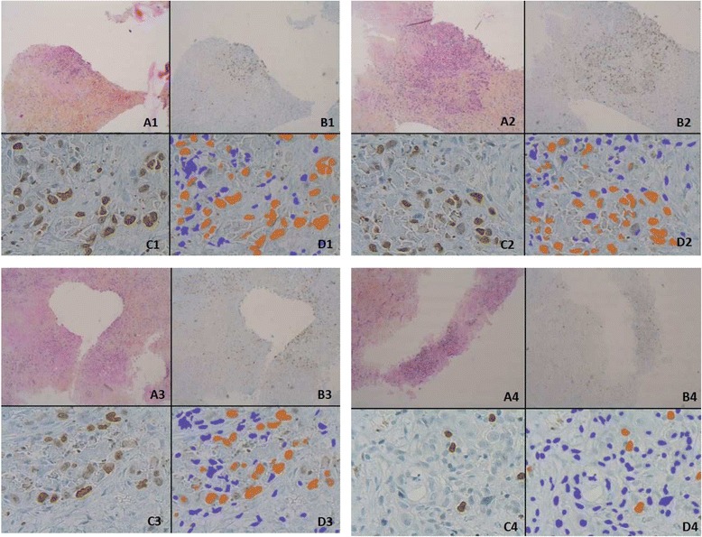
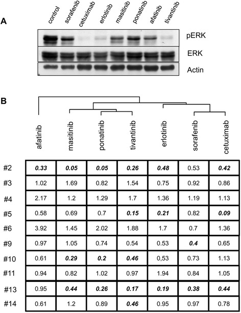
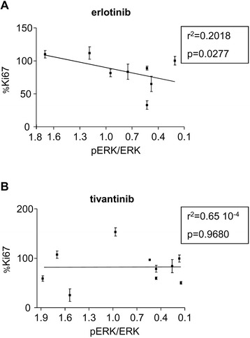
Methods: We cultivated tumour slices prepared from 18 HNSCC tumour samples obtained during surgical resection. The samples were treated for 48 h with a panel of 8 targeted therapies directed against selected oncogenic transduction pathways. We analysed the cell proliferation index (CPI) of tumour cells using Ki67 labelling and the activation status of the RAF-MEK-ERK cascade through ERK phosphorylation analysis.

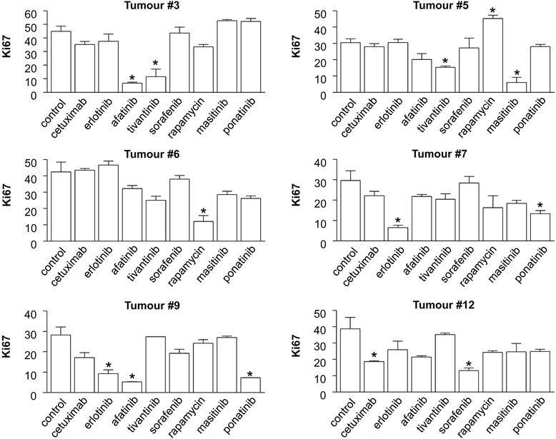
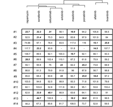
Results: Fourteen tumours were successfully analysed after short-term culture of tumour samples, revealing a striking individual heterogeneity of HNSCC in terms of tumour cell sensitivity to targeted therapies. Using 50% inhibition of CPI as threshold, sorafenib was shown to be active in 5/14 tumours. Cetuximab, the only approved targeted drug against HNSCC, was active in only 2/14 tumours. A more than 50% inhibition was observed with at least one drug out of the eight tested in 10/14 tumours. Cluster analysis was carried out in order to examine the effect of the drugs on cell proliferation and the RAF-MEK-ERK cascade.

Conclusions: In vitro culture of tumour fragments allows for the evaluation of the effects of targeted therapies on freshly resected human tumours, and might be of value as a possible guide for the design of clinical trials and for the personalization of the medical treatment of HNSCC.

Keywords: Head and neck squamous cell carcinoma; Short-term culture of tumour fragments; Targeted therapies; Treatment personalization.


Figures

Fig. 1
Fig. 1
Representative microscopic acquisitions showing tumour cell proliferation assessed by Ki67 staining. Pictures are from tumour slices obtained from four different patients and analysed after 48 h of culture. Tumour slices were maintained in control conditions (a, c) or exposed to cetuximab (b, d). Ki67 immunostaining is shown in panels a and b. Automatic identification of stained nuclei is shown in panels c and d. The nuclei that are recognized as negative for Ki67-labelling are shown in blue, while those identified as positively stained are shown in orange. The four examples shown are representative of tumour areas with different proportions of proliferating cells, as defined by the % of Ki67 labelling (X 40 in a, b; X4 in c, d)
Fig. 2
Fig. 2
Effect of targeted therapies on the cell proliferation index in six representative HNSCC tumours in short-term culture. The histograms represent the average % of tumour nuclei stained with Ki67. The average presented is based on the automatic quantification of 20 tumour fields from two slides, representing a minimum of 1000 tumour cells for each condition, as indicated in the Materials and methods section. Each treatment was maintained in culture for 48 h. Note that each individual tumour presents a different % of Ki67-positive cells in control conditions, reflecting individual differences in the basal proliferative index. * indicates a statistically-significant difference compared to control conditions, corresponding to culture without treatment, with p <0.05 (Student’s t-test)
Fig. 3
Fig. 3
Dendrogram analysis exploring the anti-proliferative effects of targeted therapies in HNSCC slices. The analysis was conducted in the 14 HNSCC tumours, based on the calculated % inhibition of cell proliferation, i.e., based on the % of labelling of Ki67 in tumour cells, after normalization. Control condition for each tumour sample was taken as reference (100 %) for the calculation of the % inhibition of Ki67-labelling by each drug. This normalization step was applied in order to minimize the impact of the heterogeneous basal proliferative index of individual tumours, and in order to preferentially address the effects of the different drugs. Following this normalization, a cluster analysis was performed in order to identify drugs with neighbouring activities on Ki67 labelling. Bold and italic characters indicate conditions with a greater than 50 % inhibition of proliferation. Empty cases indicate conditions where tumour material was not sufficient for analysis
Fig. 4
Fig. 4
ERK1/2 kinase phosphorylation analysis for the exploration of the mode of action of targeted therapies against HSNCC. a Immunoblot analysis performed upon short-term culture of a representative HNSCC tumour. Sample fragments obtained from tumour #5 were processed and analysed by immunoblotting for the indicated markers, with β-actin used as a loading control. b Dendrogram analysis based on the pERK / ERK ratio calculated for each condition. For each condition, an immunoblot analysis was performed on one slice cultured as indicated. Normalization of pERK/ERK ratio was applied in order to minimize the impact of the heterogeneity of individual tumours, by setting the value of the pERK/ERK ratio to 1 for control conditions. Following this normalization, a cluster analysis was performed in order to identify drugs with neighbouring activities on ERK phosphorylation. Bold and italic characters indicate conditions with a greater than 50 % inhibition of ERK phosphorylation
Fig. 5
Fig. 5
Correlation analysis exploring the mode of action of targeted therapies against HSNCC. A correlation is presented between the % reduction of Ki67 labelling induced by erlotinib (a) and tivantinib (b), and the % reduction of the pERK/ERK ratio. Each dot represents a single patient. Values of Pearson’s r test and the corresponding p value are presented each time

References

    1. Cancer Genome Atlas Network Comprehensive genomic characterization of head and neck squamous cell carcinomas. Nature. 2015;517:576–82. doi: 10.1038/nature14129. - DOI - PMC - PubMed
    1. Gregoire V, Lefebvre JL, Licitra L, Felip E, EHNS-ESMO-ESTRO Guidelines Working Group Squamous cell carcinoma of the head and neck: EHNS-ESMO-ESTRO Clinical Practice Guidelines for diagnosis, treatment and follow-up. Ann Oncol. 2010;21:v184–6. doi: 10.1093/annonc/mdq185. - DOI - PubMed
    1. Bonner JA, Harari PM, Giralt J, Azarnia N, Shin DM, Cohen RB, et al. Radiotherapy plus cetuximab for squamous-cell carcinoma of the head and neck. N Engl J Med. 2006;354:567–78. doi: 10.1056/NEJMoa053422. - DOI - PubMed
    1. Vermorken JB, Trigo J, Hitt R, Koralewski P, Diaz-Rubio E, Rolland F, et al. Open-label, uncontrolled, multicenter phase II study to evaluate the efficacy and toxicity of cetuximab as a single agent in patients with recurrent and/or metastatic squamous cell carcinoma of the head and neck who failed to respond to platinum-based therapy. J Clin Oncol. 2007;25:2171–7. doi: 10.1200/JCO.2006.06.7447. - DOI - PubMed
    1. Vermorken JB, Mesia R, Rivera F, Remenar E, Kawecki A, Rottey S, et al. Platinum-based chemotherapy plus cetuximab in head and neck cancer. N Engl J Med. 2008;359:1116–27. doi: 10.1056/NEJMoa0802656. - DOI - PubMed

Publication types

MeSH terms