. 2016 May 9;9(9):959-70.
doi: 10.1016/j.jcin.2016.01.032. Epub 2016 Apr 13.

Magnetic Resonance Imaging-Guided Transcatheter Cavopulmonary Shunt

Affiliations

Magnetic Resonance Imaging-Guided Transcatheter Cavopulmonary Shunt

Kanishka Ratnayaka et al. JACC Cardiovasc Interv. .

Abstract

Objectives: The aim of this study was to test the hypothesis that real-time magnetic resonance imaging (MRI) would enable closed-chest percutaneous cavopulmonary anastomosis and shunt by facilitating needle guidance along a curvilinear trajectory, around critical structures, and between a superior vena cava "donor" vessel and a pulmonary artery "target."

Background: Children with single-ventricle physiology require multiple open heart operations for palliation, including sternotomies and cardiopulmonary bypass. The reduced morbidity of a catheter-based approach would be attractive.

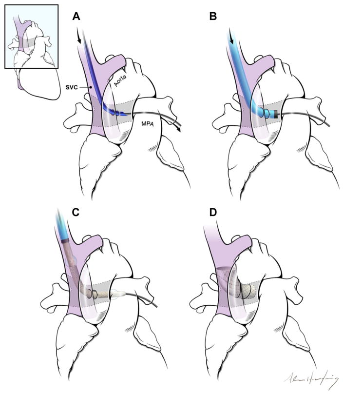
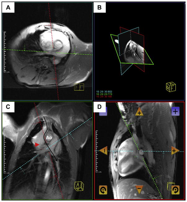
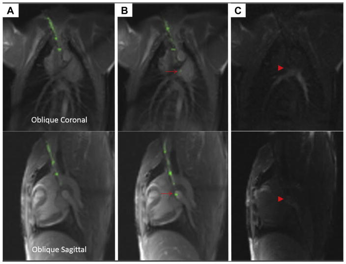
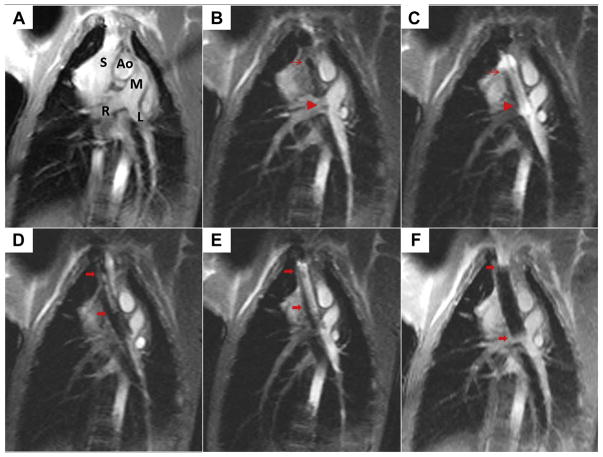
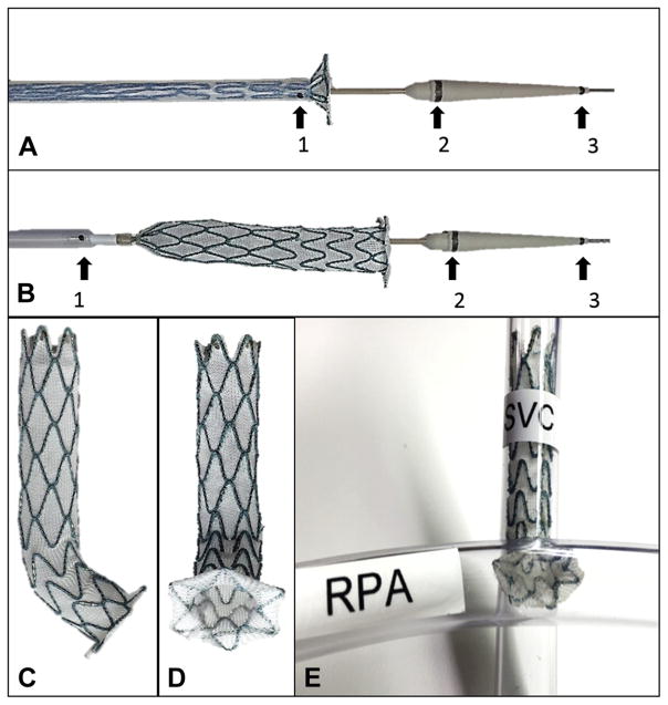
Methods: Fifteen naive swine underwent transcatheter cavopulmonary anastomosis and shunt creation under 1.5-T MRI guidance. An MRI antenna-needle was advanced from the superior vena cava into the target pulmonary artery bifurcation using real-time MRI guidance. In 10 animals, balloon-expanded off-the-shelf endografts secured a proximal end-to-end caval anastomosis and a distal end-to-side pulmonary anastomosis that preserved blood flow to both branch pulmonary arteries. In 5 animals, this was achieved with a novel, purpose-built, self-expanding device.

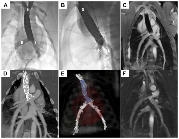
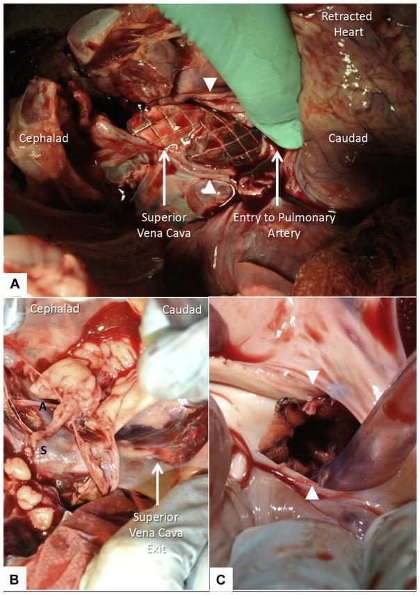
Results: Real-time MRI needle access of target vessels (pulmonary artery), endograft delivery, and superior vena cava shunt to pulmonary arteries were successful in all animals. All survived the procedure without complications. Intraprocedural real-time MRI, post-procedural MRI, x-ray angiography, computed tomography, and necropsy showed patent shunts with bidirectional pulmonary artery blood flow.

Conclusions: MRI guidance enabled a complex, closed-chest, beating-heart, pediatric, transcatheter structural heart procedure. In this study, MRI guided trajectory planning and reproducible, reliable bidirectional cavopulmonary shunt creation.

Keywords: congenital heart disease; image-guided intervention; interventional MRI; real-time MRI; sutureless anastomosis; vascular shunt.


Figures

FIGURE 1
FIGURE 1. The Concept of a Percutaneous Bidirectional Cavopulmonary Shunt
(A) First, a needle (dark blue) is advanced from the superior vena cava (SVC) (purple) into the main pulmonary artery (MPA) using magnetic resonance imaging guidance. (B) Next, the needle is exchanged for a long introducer sheath (light blue, black tip). (C) The shunt is created using endografts. (D) The endografts divert SVC blood away from the right atrium into the pulmonary arteries, while excluding the azygos vein (not shown).
FIGURE 2
FIGURE 2. Real-Time Magnetic Resonance Imaging Needle Trajectory Planning
Magnetic resonance imaging shows a typical curvilinear (C, black arrow) trajectory plan including oblique axial (A), coronal (C), sagittal (D), and 3-dimensional (B) representation of the planes, which are selected to intersect at the target pulmonary artery bifurcation. Continuous real-time imaging assists the operator in avoiding the right upper pulmonary artery branch (red arrowhead) and the aorta (Ao).
FIGURE 3
FIGURE 3. Puncturing the Pulmonary Artery Using a Magnetic Resonance Imaging Needle
This sequence depicts advancement of the magnetic resonance imaging (MRI) needle in 2 simultaneous planes, top and bottom. The MRI needle is advanced from the superior vena cava (A) and then enters the main pulmonary artery (B), where the tip is indicated with a red arrow. (C) Gadolinium contrast is injected through the needle to confirm its intraluminal position (red arrowhead).
FIGURE 4
FIGURE 4. Creating the Cavopulmonary Shunt Using Real-Time Magnetic Resonance Imaging
(A) This oblique coronal working view is shown. Ao = aorta; L = left pulmonary artery; M = main pulmonary artery; R = right pulmonary artery; S = superior vena cava. (B) The first of 2 endografts is positioned across the wall of the pulmonary artery (thin red arrow, proximal endograft; red arrowhead, distal endograft) to allow bidirectional pulmonary blood flow. (C) The first endograft is expanded using a balloon filled with gadolinium contrast. (D) The second overlapping endograft, used for want of a longer single device, is positioned to anchor cavopulmonary conduit in the superior vena cava and exclude the azygos vein (thick red arrows, proximal, distal endograft). (E) The second endograft is deployed. (F) Afterward, the completed percutaneous bidirectional cavopulmonary shunt is visible using real-time magnetic resonance imaging, where it appears black.
FIGURE 5
FIGURE 5. Novel Purpose-Built Cavopulmonary Shunt Device
A novel purpose-built, self-expanding cavopulmonary anastomosis device (A to E) and delivery (A, B) system (Transmural Systems, Andover, Massachusetts) was engineered to provide proximal end to end anastomosis (superior vena cava [SVC]) and distal end to side anastomosis (pulmonary artery) to divert superior vena cava blood flow from right atrium to both branch pulmonary arteries (E). Magnetic resonance imaging conspicuity of delivery systems was optimized with iron oxide bands (black arrows) at distal sheath tip (1), proximal nose cone (2), and distal nose cone (3). RPA = right pulmonary artery.
FIGURE 6
FIGURE 6. Novel Purpose-Built Cavopulmonary Shunt Procedure
An oblique coronal working view is used to advance the custom delivery system to target anatomy under real-time magnetic resonance imaging (MRI) guidance (A). Delivery system iron oxide marker bands are clearly seen on MRI (red arrow, distal sheath tip adjoining proximal nose cone [corresponds to 1 and 2 in Figures 5A and 5B]; red arrowhead, distal nose cone [corresponds to 3 in Figures 5A and 5B]). After precise positioning using continuous real-time MRI visualization, the novel purpose-built, self-expanding cavopulmonary anastomosis device (red arrows show proximal and distal ends of the device) is deployed (B). Gadolinium contrast injection into the superior vena cava with flow through the cavopulmonary anastomosis device into both branch pulmonary arteries confirms ideal placement (C) (Online Video 1).
FIGURE 7
FIGURE 7. The Cavopulmonary Shunt Depicted Using Radiography, Magnetic Resonance Imaging, and Computed Tomographic Angiography
X-ray contrast angiograms are shown in straight anterior-posterior (A) and straight lateral (B) projections. L = left pulmonary artery; R = right pulmonary artery; S = superior vena cava. The 3-dimensional radial steady-state free precession whole-heart magnetic resonance image (C) shows the endografts (sequential, overlapping) post-deployment. Commercial endografts are shown in A to C. Computed tomography (D, E) and steady-state free precession magnetic resonance imaging (F) show the purpose-built, self-expanding cavopulmonary anastomosis device post-deployment.
FIGURE 8
FIGURE 8. Necropsy
At necropsy, the walls of the superior vena cava (white arrowheads) and the rest of the heart are retracted manually to expose tandem overlapping endografts bypassing superior vena cava blood flow to pulmonary arteries (A). The endograft exits the posterior wall of the superior vena cava caudad and re-enters the pulmonary artery. Similarly, the novel, purpose-built, self-expanding cavopulmonary endograft (B, C) bypasses superior vena cava blood flow to both branch pulmonary arteries. A = azygous vein; S = superior vena cava. The walls of the pulmonary artery (white arrowheads) are retracted manually (C) to expose distal end-to-side anastomosis of the novel endograft.

Comment in

References

    1. Glenn WW. Circulatory bypass of the right side of the heart. IV. Shunt between superior vena cava and distal right pulmonary artery; report of clinical application. N Engl J Med. 1958;259:117–20. - PubMed
    1. Hopkins RA, Armstrong BE, Serwer GA, Peterson RJ, Oldham HN., Jr Physiological rationale for a bidirectional cavopulmonary shunt. A versatile complement to the Fontan principle. J Thorac Cardiovasc Surg. 1985;90:391–8. - PubMed
    1. Saikus CE, Ratnayaka K, Barbash IM, et al. MRI-guided vascular access with an active visualization needle. J Magn Reson Imaging. 2011;34:1159–66. - PMC - PubMed
    1. Ratnayaka K, Faranesh AZ, Hansen MS, et al. Real-time MRI-guided right heart catheterization in adults using passive catheters. Eur Heart J. 2013;34:380–9. - PMC - PubMed
    1. Pandya B, Quail MA, Steeden JA, et al. Real-time magnetic resonance assessment of septal curvature accurately tracks acute hemodynamic changes in pediatric pulmonary hypertension. Circ Cardiovasc Imaging. 2014;7:706–13. - PubMed

Publication types

LinkOut - more resources