Review
. 2016 Jun;7(3):341-50.
doi: 10.1007/s13244-016-0490-9. Epub 2016 Apr 16.

Post traumatic deafness: a pictorial review of CT and MRI findings

Affiliations
Review

Post traumatic deafness: a pictorial review of CT and MRI findings

Olivier Maillot et al. Insights Imaging. 2016 Jun.

Abstract

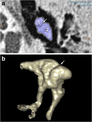
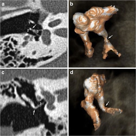
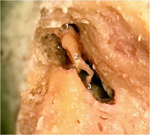
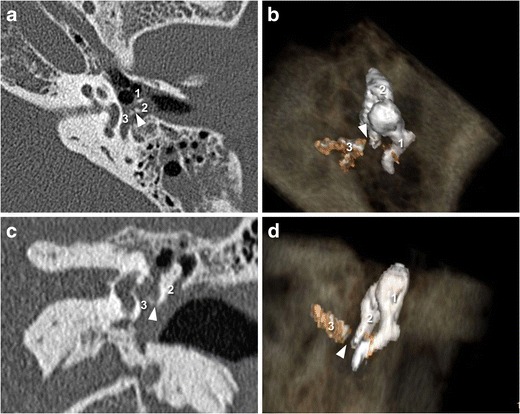
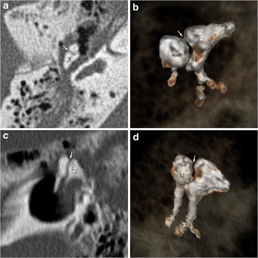
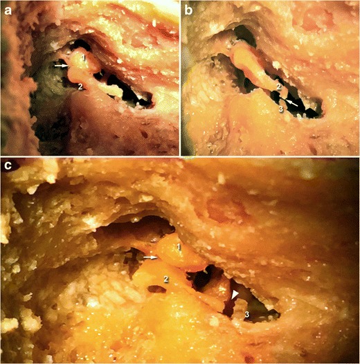
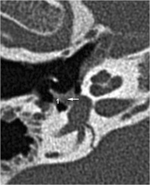
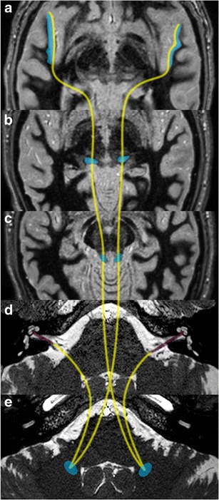
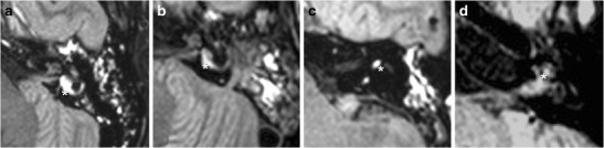
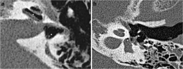
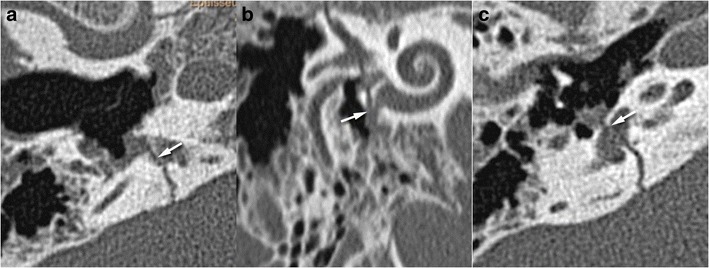
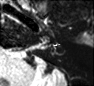
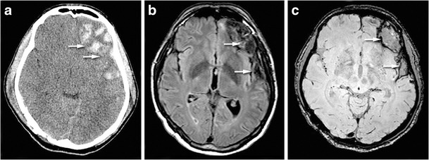
Hearing loss is a common functional disorder after trauma, and radiologists should be aware of the ossicular, labyrinthine or brain lesions that may be responsible. After a trauma, use of a systematic approach to explore the main functional components of auditory pathways is essential. Conductive hearing loss is caused by the disruption of the conductive chain, which may be due to ossicular luxation or fracture. This pictorial review firstly describes the normal 2-D and 3-D anatomy of the ossicular chain, including the incudo-malleolar and incudo-stapedial joints. The role of 3-D CT in the post-traumatic evaluation of injury to the temporal bone is then evaluated. In the case of sensorineural hearing loss, CT can detect pneumolabyrinth and signs of perilymphatic fistulae but fails to detect subtle lesions within the inner ear, such as labyrinthine haemorrhage or localized brain axonal damage along central auditory pathways. The role that MRI with 3-D-FLAIR acquisition plays in the detection of inner ear haemorrhage and post-traumatic lesions of the brain parenchyma that may lead to auditory agnosia is also discussed.

Key points: • The most common middle ear injuries are incudo-malleolar and incudo-stapedial joint luxation. • In patients with SNHL, CT can detect pneumolabyrinth or perilymphatic fistula • 3-D-FLAIR MRI appears the best sequence to highlight labyrinthine haemorrhage • Axonal damage and brain hematoma may lead to deafness.

Keywords: CT scan; Ear ossicles; Magnetic resonance imaging; Temporal bone deafness; Trauma.


Figures

Fig. 1
Fig. 1
2-D CT images in the axial plane showing a normal incudo-malleolar joint. The ice-cream cone including the body of the incus (2) and the head of the malleus (1) 3-D CT reveals that malleus (1) and incus (2) are in close contact in the healthy middle ear
Fig. 2
Fig. 2
2-D CT images in the axial and oblique planes showing a normal incudo-stapedial joint. The malleus is annotated (1) The axial and oblique planes (a and c) show the ossicular « V », comprising the long process of the incus (2) and the stapes (3). 3-D-CT acquisition (b and d) reveal continuity between the stapes head and the long process of the incus in the healthy middle ear
Fig. 3
Fig. 3
Cadaveric view showing a normal incudo-malleolar joint on the left side and a normal incudo-stapedial joint on the right side. As on the 3-D CT views, the malleus (1) and the incus (2) are in close contact. The incus is also in continuity with the stapes (3)
Fig. 4
Fig. 4
2-D CT images in the axial and oblique planes (a and c) showing a post-traumatic incudo-stapedial joint luxation. The malleus is annotated (1) The oblique plane shows disruption of the ossicular « V », comprising the stapes (3) and the long process of the incus (2). The 3-D CT images (b and d) also reveal the “gap” (white arrowhead) between the stapes (3) and the incus (2)
Fig. 5
Fig. 5
2-D-CT images (a and c) in the axial and coronal planes showing the incudo-malleolar joint after temporal bone trauma. The incus (2) is no longer in contact with the head of the malleus (1) on 2-D and 3-D-CT acquisition (b and d)
Fig. 6
Fig. 6
Cadaveric view showing incudo-malleolar luxation (white arrow, upper left) and incudo-stapedial luxation (white arrow, upper right). On the third image, both joints are dislocated (white arrow and white arrowhead). The malleus is annotated (1), the incus (2), and the stapes (3)
Fig. 7
Fig. 7
Stapedio-vestibular luxation: CT stapes view highlighting stapedio-vestibular luxation. The perilymphatic liquid leaking through the oval window (1) implies rupture of the annular ligament
Fig. 8
Fig. 8
Central auditory pathways: the auditory cortex is located in the superior temporal gyrus (a: blue areas). Beyond the cochlear nerves (d: coloured in purple); the central auditory pathways run through the cochlear nuclei, the inferior colliculii (c) before they decussate to the contralateral olivary nucleus (e), run through the medial geniculate body (b) and finally reach the auditory radiations
Fig. 9
Fig. 9
Examples of inner ear haemorrhage: the inner ear haemorrhage (white star) appears as a hypersignal on nonenhanced FLAIR acquired images of the vestibule (a and b), superior semicircular canal (c), and the cochlea (d). The first and third patient (a and c) referred with trans-labyrinthine fracture, the second (b) with extra-labyrinthine fracture, and the fourth (d) with major brain trauma, without temporal bone fracture. All patients presented post-traumatic SNHL, ipsilateral to the haemorrhage
Fig. 10
Fig. 10
a Perilymphatic fistulae: CT axial views showing a perilymphatic fistula, highlighted by a pneumolabyrinth (white stars). Perilymphatic liquid has leaked into the middle ear. Massive pneumolabyrinth was seen 1 month after a translabyrinthine fracture, with air in the perilymphatic space (scala vestibuli and the vestibule) b Example of pneumolabyrinth without temporal bone fracture (white star)
Fig. 11
Fig. 11
Perilymphatic fistulae: round and oval windows are the two communication routes between the middle ear and inner ear, implying two weakness zones. Axial and oblique CT views showing fractures (white arrows) crossing the round (a, b) and oval (c) windows
Fig. 12
Fig. 12
Post-traumatic endolymphatic hydrops: for this patient with extra-labyrinthine fracture and post-traumatic left SNHL, the vestibular hydrops was obvious with an endolymphatic vestibular area (white arrow), encompassing up to 50 % of the whole vestibular surface. However, the role of trauma as a direct cause of the endolymphatic hydrops remains unclear
Fig. 13
Fig. 13
Brain haemorrhage in a patient with contralateral auditory agnosia. CT view (a) showing left fronto-temporal hemorrhagic injury. FLAIR sequence (b) and susceptibility-weighted imaging (c) showing the cortical damage in particular in the superior temporal gyrus and in the basifrontal region (white arrows)

References

    1. Sun GH, Shoman NM, Samy RN, et al. Do contemporary temporal bone fracture classification systems reflect concurrent intracranial and cervical spine injuries? Laryngoscope. 2011;121:929–932. doi: 10.1002/lary.21718. - DOI - PubMed
    1. Cvorovic L, Ljiljana C, Jovanovic MB, et al. Management of complication from temporal bone fractures. Eur Arch Otorhinolaryngol. 2012;269:399–403. doi: 10.1007/s00405-011-1692-x. - DOI - PubMed
    1. Fatterpekar GM, Doshi AH, Dugar M, et al. Role of 3D CT in the evaluation of the temporal bone. Radiographics. 2006;26(Suppl 1):S117–S132. doi: 10.1148/rg.26si065502. - DOI - PubMed
    1. Leng S, Diehn FE, Lane JI, et al. Temporal bone CT: improved image quality and potential for decreased radiation dose using an ultra-high-resolution scan mode with an iterative reconstruction algorithm. AJNR Am J Neuroradiol. 2015;36:1599–1603. doi: 10.3174/ajnr.A4338. - DOI - PMC - PubMed
    1. Mark AS, Seltzer S, Harnsberger HR. Sensorineural hearing loss: more than meets the eye? AJNR Am J Neuroradiol. 1993;14:37–45. - PMC - PubMed

LinkOut - more resources