

Programmed death-ligand 1 overexpression is a prognostic marker for aggressive papillary thyroid cancer and its variants
- PMID: 27086918
- PMCID: PMC5078015
- DOI: 10.18632/oncotarget.8698
Programmed death-ligand 1 overexpression is a prognostic marker for aggressive papillary thyroid cancer and its variants
Abstract
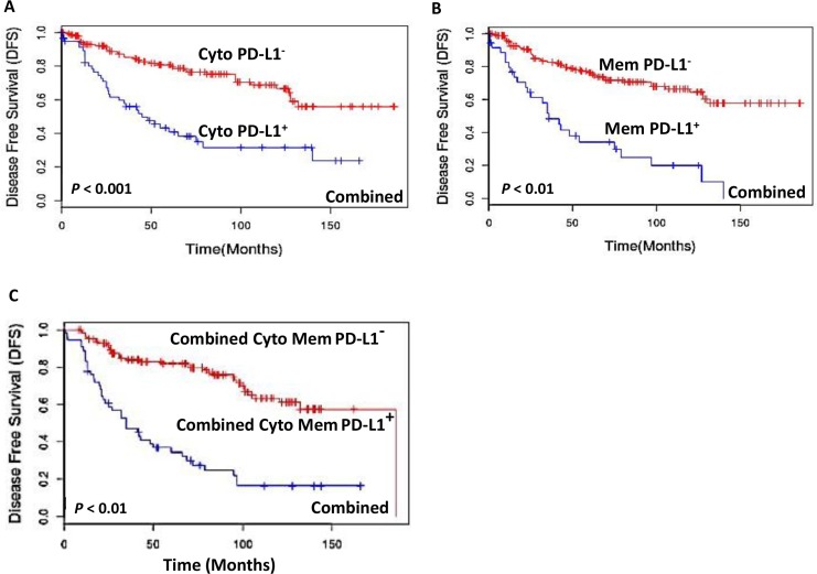
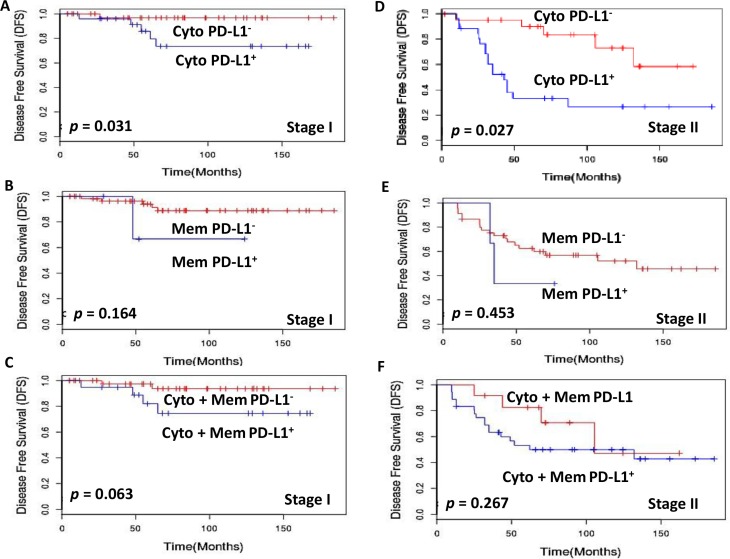
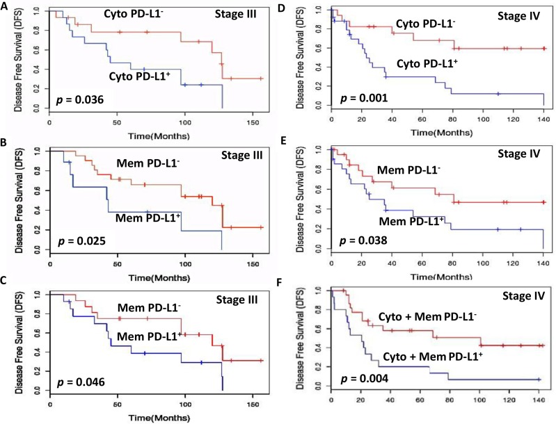
Programmed death-ligand 1(PD-L1) expression on tumor cells is emerging as a potential predictive biomarker in anti-PD-L1 directed cancer immunotherapy. We analyzed PD-L1 expression in papillary thyroid carcinoma (PTC) and its variants and determined its prognostic potential to predict clinical outcome in these patients. This study was conducted at an academic oncology hospital which is a prime referral centre for thyroid diseases. Immunohistochemical subcellular localization (IHC) analyses of PD-L1 protein was retrospectively performed on 251 archived formalin fixed and paraffin embedded (FFPE) surgical tissues (66 benign thyroid nodules and 185 PTCs) using a rabbit monoclonal anti-PD-L1 antibody (E1L3N, Cell Signaling Technology) and detected using VECTASTAIN rapid protocol with diaminobenzidine (DAB) as the chromogen. The clinical-pathological factors and disease outcome over 190 months were assessed; immunohistochemical subcellular localization of PD-L1 was correlated with disease free survival (DFS) using Kaplan Meier survival and Cox multivariate regression analysis. Increased PD-L1 immunostaining was predominantly localized in cytoplasm and occasionally in plasma membrane of tumor cells. Among all combined stages of PTC, patients with increased PD-L1 membrane or cytoplasmic positivity had significantly shorter median DFS (36 months and 49 months respectively) as compared to those with PD-L1 negative tumors (DFS, both 186 months with p < 0.001 and p < 0.01 respectively). Comparison of PD-L1+ and PD-L1- patients with matched staging showed increased cytoplasmic positivity in all four stages of PTC that correlated with a greater risk of recurrence and a poor prognosis, but increased membrane positivity significantly correlated with a greater risk of metastasis or death only in Stage IV patients. In conclusion, PD-L1 positive expression in PTC correlates with a greater risk of recurrence and shortened disease free survival supporting its potential application as a prognostic marker for PTC.
Keywords: benign nodule; programmed death-ligand 1; protein biomarkers; subcellular localization; thyroid cancer.
Conflict of interest statement
PGW and RR are shareholders in Proteocyte Diagnostics Inc. All the other authors (SC, JV, FJ, OP, AA and CM) have nothing to disclose.
Figures

Similar articles
-
Programmed Death-Ligand 1 Expression in Papillary Thyroid Cancer and Its Correlation with Clinicopathologic Factors and Recurrence.Thyroid. 2017 Apr;27(4):537-545. doi: 10.1089/thy.2016.0228. Epub 2017 Jan 24. Thyroid. 2017. PMID: 27825291
-
Independent prognostic role of PD-L1expression in patients with esophageal squamous cell carcinoma.Oncotarget. 2017 Jan 31;8(5):8315-8329. doi: 10.18632/oncotarget.14174. Oncotarget. 2017. PMID: 28039448 Free PMC article.
-
Programmed Death - Ligand 1 Expression Distinguishes Invasive Encapsulated Follicular Variant of Papillary Thyroid Carcinoma from Noninvasive Follicular Thyroid Neoplasm with Papillary-like Nuclear Features.EBioMedicine. 2017 Apr;18:50-55. doi: 10.1016/j.ebiom.2017.03.031. Epub 2017 Mar 24. EBioMedicine. 2017. PMID: 28363612 Free PMC article.
-
Programmed Death-Ligand 1 (PD-L1) Is a Potential Biomarker of Disease-Free Survival in Papillary Thyroid Carcinoma: a Systematic Review and Meta-Analysis of PD-L1 Immunoexpression in Follicular Epithelial Derived Thyroid Carcinoma.Endocr Pathol. 2020 Sep;31(3):291-300. doi: 10.1007/s12022-020-09630-5. Endocr Pathol. 2020. PMID: 32468210
-
The clinicopathological and prognostic significance of PD-L1 expression in pancreatic cancer: A meta-analysis.Hepatobiliary Pancreat Dis Int. 2018 Apr;17(2):95-100. doi: 10.1016/j.hbpd.2018.03.007. Epub 2018 Mar 13. Hepatobiliary Pancreat Dis Int. 2018. PMID: 29576277 Review.
Cited by
-
Peripheral Versus Intraparenchymal Papillary Thyroid Microcarcinoma: Different Morphologies and PD-L1 Expression.Head Neck Pathol. 2022 Mar;16(1):200-212. doi: 10.1007/s12105-021-01337-1. Epub 2021 Jun 2. Head Neck Pathol. 2022. PMID: 34076845 Free PMC article.
-
How cancer cells make and respond to interferon-I.Trends Cancer. 2023 Jan;9(1):83-92. doi: 10.1016/j.trecan.2022.09.003. Epub 2022 Oct 8. Trends Cancer. 2023. PMID: 36216730 Free PMC article. Review.
-
Unfolding of Imminent Bio-Signatures in the Prognosis of Thyroid Cancer; The Emergence of Estrogen Related Receptor Gamma (ERRγ) as a Hurricane.Asian Pac J Cancer Prev. 2023 Feb 1;24(2):375-387. doi: 10.31557/APJCP.2023.24.2.375. Asian Pac J Cancer Prev. 2023. PMID: 36853284 Free PMC article. Review.
-
Molecular Alterations in Thyroid Cancer: From Bench to Clinical Practice.Genes (Basel). 2019 Sep 13;10(9):709. doi: 10.3390/genes10090709. Genes (Basel). 2019. PMID: 31540307 Free PMC article. Review.
-
HIF1α/PD-L1 axis mediates hypoxia-induced cell apoptosis and tumor progression in follicular thyroid carcinoma.Onco Targets Ther. 2019 Aug 13;12:6461-6470. doi: 10.2147/OTT.S203724. eCollection 2019. Onco Targets Ther. 2019. Retraction in: Onco Targets Ther. 2022 Nov 21;15:1403-1404. doi: 10.2147/OTT.S397860. PMID: 31616157 Free PMC article. Retracted.
References
-
- Sipos JA, Mazzaferri EL. Thyroid cancer epidemiology and prognostic variables. Clin Oncol (R Coll Radiol) 2010;22:395–404. - PubMed
-
- Bethesda, MD: National Institute of Health; http://seer.cancer.gov/statfacts/html/thyro.htmlNational Cancer Institute 2015 Cancer Statistics.http://www.cancer.gov/statistics National Cancer institute.
-
- O'Neill JP, Power D, Condron C, Bouchier-Hayes D, Walsh M. Anaplastic thyroid cancer, tumorigenesis and therapy. Ir J Med Sci. 2010;179:9–15. - PubMed
-
- Green LD, Mack L, Pasieka JL. Anaplastic thyroid cancer and primary thyroid lymphoma: a review of these rare thyroid malignancies. J Surg Oncol. 2006;94:725–736. - PubMed
-
- Mazzaferri EL, Jhiang SM. Long-term impact of initial surgical and medical therapy on papillary and follicular thyroid cancer. Am J Med. 1994;97:418–428. - PubMed
Publication types
MeSH terms
Substances
LinkOut - more resources
Full Text Sources
Other Literature Sources
Medical
Research Materials
