. 2016 Apr 18:6:24436.
doi: 10.1038/srep24436.

Lymphatic transport of exosomes as a rapid route of information dissemination to the lymph node

Affiliations

Lymphatic transport of exosomes as a rapid route of information dissemination to the lymph node

Swetha Srinivasan et al. Sci Rep. .

Abstract

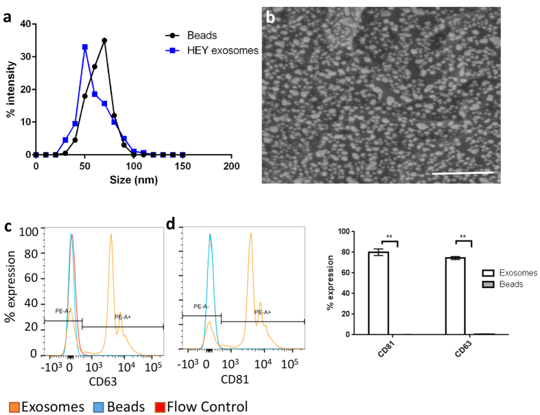
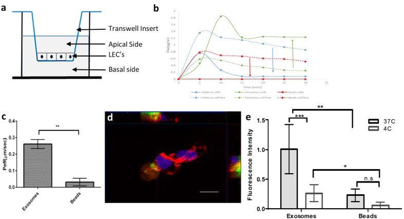
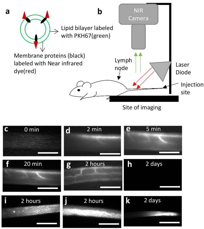
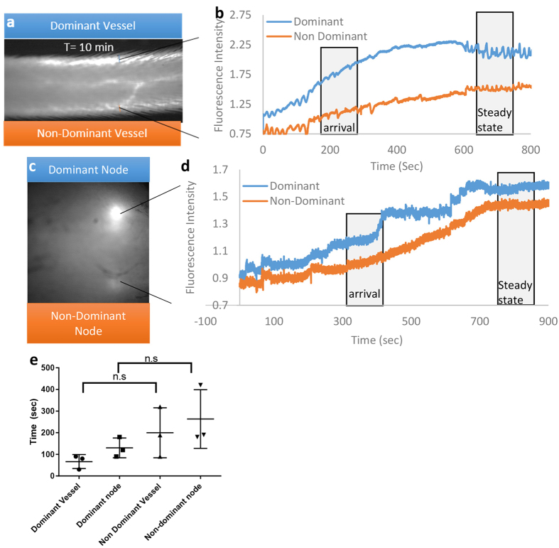
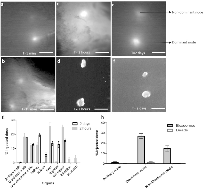
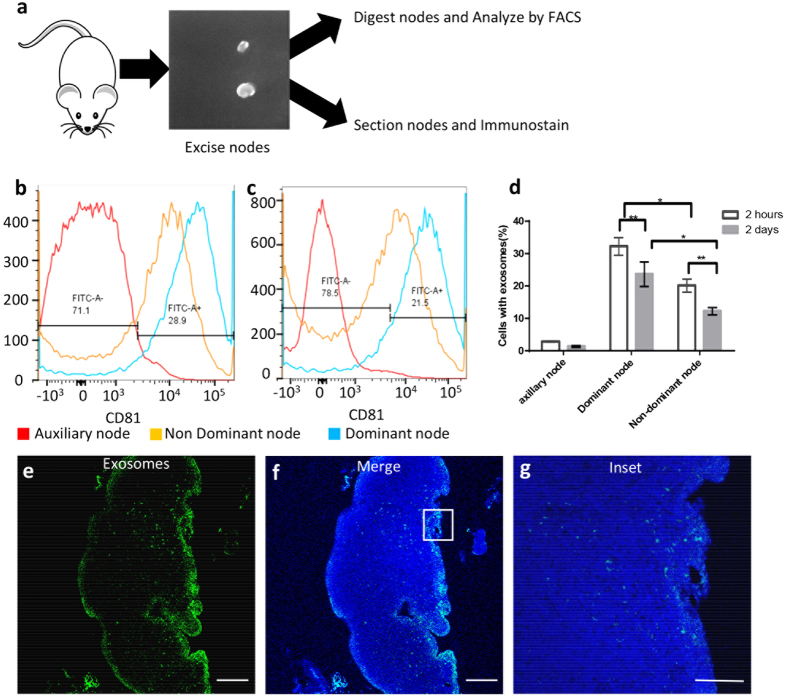
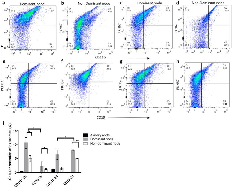
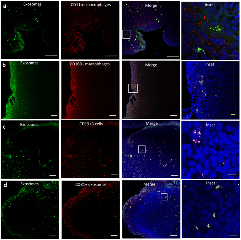
It is well documented that cells secrete exosomes, which can transfer biomolecules that impact recipient cells' functionality in a variety of physiologic and disease processes. The role of lymphatic drainage and transport of exosomes is as yet unknown, although the lymphatics play critical roles in immunity and exosomes are in the ideal size-range for lymphatic transport. Through in vivo near-infrared (NIR) imaging we have shown that exosomes are rapidly transported within minutes from the periphery to the lymph node by lymphatics. Using an in vitro model of lymphatic uptake, we have shown that lymphatic endothelial cells actively enhanced lymphatic uptake and transport of exosomes to the luminal side of the vessel. Furthermore, we have demonstrated a differential distribution of exosomes in the draining lymph nodes that is dependent on the lymphatic flow. Lastly, through endpoint analysis of cellular distribution of exosomes in the node, we identified macrophages and B-cells as key players in exosome uptake. Together these results suggest that exosome transfer by lymphatic flow from the periphery to the lymph node could provide a mechanism for rapid exchange of infection-specific information that precedes the arrival of migrating cells, thus priming the node for a more effective immune response.


Figures

Figure 1
Figure 1. Characterization of exosomes and beads.
(a) Size distribution of HEY exosomes as compared to that of beads. (b) Scanning electron micrograph of exosomes. Scale bar = 500 nm. (c) Expression of CD63 and (d) CD81 on exosomes and beads. (e) Quantitation of CD63 and CD81 on exosomes and beads by flow cytometry (p-value < 0.01).
Figure 2
Figure 2. Exosomes transported rapidly and selectively through the lymphatic endothelium in vitro.
(a) Schematic of transport experiment, (b) Transport of exosomes across the lymphatic endothelium occurs rapidly (t = 5–30 mins) and is enhanced in the presence of cells, (c) Exosomes are selectively transported into the lymphatic endothelium (versus beads), (d) Orthogonal view of LEC’s (nuclei stained with DAPI, actin stained red) with PKH67 exosomes and at 37 °C. Scale bar, 5 μm and (e) quantitation of exosome and beads in cells by fluorescence intensity at 37 °C and 4 °C.
Figure 3
Figure 3. Exosomes are transported rapidly through the lymphatic endothelium in vivo.
(a) Dual labeling of exosomes, (b) injection and visualization scheme in mice. Exosomes are detected in the lymphatics rapidly (c) vessel at 0 mins, (d) vessel at 2 mins, (e) vessel at 5 mins, (f) vessel at 20 mins (g) vessel at 2 hours, (h) vessel at 2 days, (i) lymphatic capillaries seen close to the injection site at 2 hours, (j) injection site at 2 hours, and (k) injection site at 2 days. Scale bar; 5 mm.
Figure 4
Figure 4. Characterization of exosomes transport in vivo.
(a) Steady state fluorescence in the lymphatic collecting vessel (b) Intensity profile of a specified region of interest of exosome transport in a representative vessel over a 10 minute period, (c) Steady state fluorescence in the draining lymph node, (d) Intensity profile of a specified region of interest of exosome transport in a representative lymph node over a 10 minute period, (e) Arrival time of detectable levels of fluorescence for dominant and non-dominant collecting vessels and draining lymph nodes.
Figure 5
Figure 5. Characterization of exosome retention in vivo.
Exosomes are detected in the node rapidly: (a) Only the dominant node is visible at 5 mins in vivo, (b) Both nodes are visible at 15 mins in vivo, (c) Draining lymph nodes visualized at 2 h pre-excision (in animal), (d) Excised lymph nodes at 2 h post injection, (e) Draining lymph nodes visualized at 2d pre-excision (in animal) (f) Excised lymph nodes at 2 days post injection, Scale = 5 mm. (g) Biodistribution of exosomes in mice organs analyzed at 2 hours and 2 days post injection and (h) quantitation of exosomes and beads retained in the lymph node 1 hour post injection as determined by fluorescence.
Figure 6
Figure 6. Characterization of exosome retention in the draining lymph node.
(a) Schematic of node procession post excision from mouse (b) Dominant node retains a larger quantity of exosomes at 2 hours (c) Dominant node retains a larger quantity of exosomes at 2 days, (d) Quantitation of exosome retention by the dominant and non-dominant nodes at 2 hours and 2 days respectively, (e) Exosome localization within the node at 2 days; (f) Merged image with whole node nuclear staining and exosome localization, (g) magnified area in the node showing exosome localization. Scale bar; 10 um.
Figure 7
Figure 7. Characterization of exosome uptake by CD11b and CD19 cells in the node by flow cytometry.
The dominant node was digested and stained for (a) CD11b at 2 h, (c) CD11b at 2 days, (e) CD19 at 2 hours, (g) CD19 at 2 days. The non-dominant node was stained for (b) CD11b at 2 h, (d) CD11b at 2 days, (f) CD19 at 2 hours, (h) CD19 at 2 days and (i) quantitation of exosome uptake by the dominant and non-dominant nodes at 2 hours and 2 days respectively expressed as a percentage of PKH67 positive cells.
Figure 8
Figure 8. Localization of exosomes within the lymph node.
Shown are serial lymph node sections at 2 days following injection of 10 ug of exosomes (green). Immune cells were identified as indicated (red) with antibodies against (a) CD11b (macrophages), (b) CD169 (macrophages), and (c) CD19 (B-cells), (d) CD81(red) was used as a secondary localization marker to confirm exosome retention in the node. White scale bar = 50 um while yellow scale bar is 5 um.

References

    1. Loo Y. M. & Gale M. Jr. Immune signaling by RIG-I-like receptors. Immunity 34, 680–692, doi: 10.1016/j.immuni.2011.05.003 (2011). - DOI - PMC - PubMed
    1. Kawai T. & Akira S. Toll-like receptors and their crosstalk with other innate receptors in infection and immunity. Immunity 34, 637–650, doi: 10.1016/j.immuni.2011.05.006 (2011). - DOI - PubMed
    1. Janeway C. A. Jr. & Medzhitov R. Innate immune recognition. Annual review of immunology 20, 197–216, doi: 10.1146/annurev.immunol.20.083001.084359 (2002). - DOI - PubMed
    1. Thery C., Zitvogel L. & Amigorena S. Exosomes: composition, biogenesis and function. Nature reviews. Immunology 2, 569–579, doi: 10.1038/nri855 (2002). - DOI - PubMed
    1. Guermonprez P., Valladeau J., Zitvogel L., Théry C. & Amigorena S. Antigen presentation and t cell stimulation by dendritic cells. Annu Rev Immunol 20, 621–667, doi: 10.1146/annurev.immunol.20.100301.064828 (2002). - DOI - PubMed

Publication types