. 2016 Jun;233(12):2383-97.
doi: 10.1007/s00213-016-4289-7. Epub 2016 Apr 18.

The adenosinergic system is involved in sensitization to morphine withdrawal signs in rats-neurochemical and molecular basis in dopaminergic system

Affiliations

The adenosinergic system is involved in sensitization to morphine withdrawal signs in rats-neurochemical and molecular basis in dopaminergic system

Joanna Listos et al. Psychopharmacology (Berl). 2016 Jun.

Abstract

Rationale: Experimental data informs that not only do the dose and time duration of dependent drugs affect the severity of withdrawal episodes. Previous withdrawal experiences may intensify this process, which is referred as sensitization to withdrawal signs. Adenosine and dopamine (DA) receptors may be involved in this sensitization.

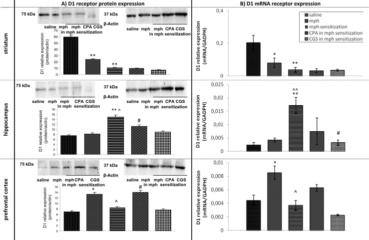
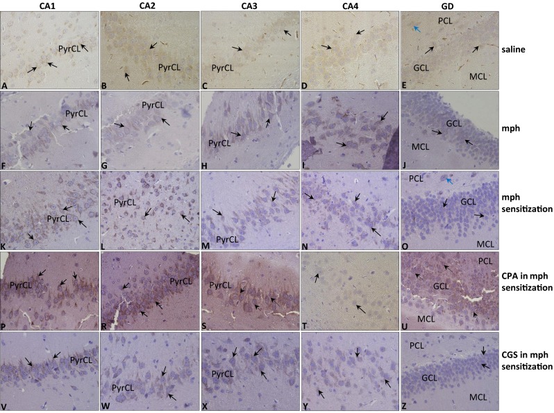
Objectives: Rats were continuously and sporadically treated with increasing doses of morphine for 8 days. In rats, sporadically treated with morphine, morphine administration was modified by adding three morphine-free periods. Adenosine agonists were given during each of the morphine-free periods (six injections in total). On the 9th day, morphine was injected. One hour later, naloxone was administered to induce morphine withdrawal signs. Then, the animals were placed into cylinders and the number of jumpings was recorded. Next, the rats were decapitated and brain and brain structures (striatum, hippocampus, and prefrontal cortex) were dissected for neurochemical, molecular, and immunohistochemical experiments within DAergic pathways.

Results: We demonstrated that previous experiences of opioid withdrawal intensified subsequent withdrawal signs. Adenosine ligands attenuated the sensitization to withdrawal signs. In a neurochemical study, the release of DA and its metabolites was impaired in all structures. Significant alterations were also observed in mRNA and protein expression of DA receptors.

Conclusions: Results demonstrate that intermittent treatment with morphine induces alterations in the DAergic system which may be responsible for sensitization to morphine withdrawal signs. Although adenosine ligands attenuate this type of sensitization, they are not able to fully restore the physiological brain status.

Keywords: Dependence; Dopamine receptor expression; Morphine withdrawal signs; Naloxone.


Figures

Scheme 1
Scheme 1
Shows the procedure of administration of morphine in morphine group and morphine and adenosine agonists (CPA and CGS 21680) in morphine-sensitized group
Fig. 1
Fig. 1
Effects of continuous and sporadic treatment with increasing doses of morphine on the intensity of naloxone-induced morphine (mph) withdrawal signs (jumpings). The role of adenosine A1 and A2A agonists. Rats were treated in two schemes: for 8 continuous days (mph group) and for four 2-day periods (4 × 2) interspersed with morphine-free period days (mph-sensitized group). Adenosine agonists (CPA—0.1 mg/kg, ip and CGS 21680—0.1 mg/kg, ip) were administered during the morphine-free periods. Naloxone (2.0 mg/kg, ip) was administered for induction of morphine withdrawal signs. *p < 0.05, ***p < 0.001 vs saline group, ^^^p < 0.001 vs mph group, ### p < 0.001 vs mph-sensitized group (Tukey’s test)
Fig. 2
Fig. 2
Representative western blots and densitometric analysis of protein (normalized to β-actin) (a) and densitometric analysis mRNA levels of D1 receptor (b) in brain of continuously and sporadically treated with morphine (mph) rats. The role of adenosine A1 and A2A agonists. The results are expressed as means ± SD from different areas of three to four rat brains. *p < 0.05, **p < 0.01 vs saline group; ^ p < 0.05, ^^ p < 0.01 vs mph group; # p < 0.05 vs mph-sensitized group (Mann–Whitney test)
Fig. 3
Fig. 3
Representative western blots and densitometric analysis of protein (normalized to β-actin) (a) and densitometric analysis mRNA levels of D2 receptor (b) in brain of continuously and sporadically treated with morphine (mph) rats. The role of adenosine A1 and A2A agonists. The results are expressed as means ± SD from different areas of three to four rat brains. *p < 0.05, **p < 0.01, ***p < 0.001 vs saline group; ^ p < 0.05, ^^ p < 0.01 vs mph group; # p < 0.05, ## p < 0.01, ### p < 0.001 vs mph-sensitized group (Mann–Whitney test)
Fig. 4
Fig. 4
Immunolocalization of D1 receptor in hippocampus of rats during withdrawal period induced by naloxone in: saline group (AE); morphine group (mph) (FJ); morphine-sensitized group (mph sensitization) (KO); CPA in mph-sensitized group (PU); and CGS in mph-sensitized group (VZ); objective magnification ×40. (CA1–CA4) area of (CA) cornu ammonis; (GD) gyrus dentate; (PyrCL) pyramidal cell layer; (GCL) granular cell layer; (PCL) polymorphic cell layer; (MCL) molecular cell layer
Fig. 5
Fig. 5
Immunolocalization of D2 receptor in hippocampus of rats during withdrawal period induced by naloxone in: saline group (AE); morphine group (mph) (F–J); morphine-sensitized group (mph sensitization) (KO); CPA in mph-sensitized group (PU); and CGS in mph-sensitized group (VZ); objective magnification ×40. (CA1–CA4) area of (CA) cornu ammonis; (GD) gyrus dentate; (PyrCL) pyramidal cell layer; (GCL) granular cell layer; (PCL) polymorphic cell layer; (MCL) molecular cell layer

References

    1. Acquas E, Carboni E, Di Chiara G. Profound depression of mesolimbic DA release after morphine withdrawal in dependent rats. Eur J Pharmacol. 1991;193:133–134. doi: 10.1016/0014-2999(91)90214-B. - DOI - PubMed
    1. Ahlijanian MK, Takemori AE. Changes in adenosine receptor sensitivity in morphine-tolerant and -dependent mice. J Pharmacol Exp Ther. 1986;236:615–620. - PubMed
    1. Baldo BA, Koob GF, Markou A. Role of adenosine A2 receptors in brain stimulation reward under baseline conditions and during cocaine withdrawal in rats. J Neurosci. 1999;19:11017–11026. - PMC - PubMed
    1. Balter RE, Dykstra L. The effect of environmental factors on morphine withdrawal in C57BL/6J mice: running wheel access and group housing. Psychopharmacology. 2012;224:91–100. doi: 10.1007/s00213-012-2826-6. - DOI - PubMed
    1. Bassareo V, Tanda G, Di Chiara G. Increase of extracellular DA in the medial prefrontal cortex during spontaneous and naloxone-precipitated opiate abstinence. Psychopharmacology. 1995;122:202–205. doi: 10.1007/BF02246097. - DOI - PubMed

LinkOut - more resources