. 2016:2016:5184601.
doi: 10.1155/2016/5184601. Epub 2016 Feb 10.

Phenotypic and Functional Characterization of Mesenchymal Stem/Multipotent Stromal Cells from Decidua Basalis of Human Term Placenta

Affiliations

Phenotypic and Functional Characterization of Mesenchymal Stem/Multipotent Stromal Cells from Decidua Basalis of Human Term Placenta

F M Abomaray et al. Stem Cells Int. 2016.

Abstract

Mesenchymal stem cell (MSC) therapies for the treatment of diseases associated with inflammation and oxidative stress employ primarily bone marrow MSCs (BMMSCs) and other MSC types such as MSC from the chorionic villi of human term placentae (pMSCs). These MSCs are not derived from microenvironments associated with inflammation and oxidative stress, unlike MSCs from the decidua basalis of the human term placenta (DBMSCs). DBMSCs were isolated and then extensively characterized. Differentiation of DBMSCs into three mesenchymal lineages (adipocytes, osteocytes, and chondrocytes) was performed. Real-time polymerase chain reaction (PCR) and flow cytometry techniques were also used to characterize the gene and protein expression profiles of DBMSCs, respectively. In addition, sandwich enzyme-linked immunosorbent assay (ELISA) was performed to detect proteins secreted by DBMSCs. Finally, the migration and proliferation abilities of DBMSCs were also determined. DBMSCs were positive for MSC markers and HLA-ABC. DBMSCs were negative for hematopoietic and endothelial markers, costimulatory molecules, and HLA-DR. Functionally, DBMSCs differentiated into three mesenchymal lineages, proliferated, and migrated in response to a number of stimuli. Most importantly, these cells express and secrete a distinct combination of cytokines, growth factors, and immune molecules that reflect their unique microenvironment. Therefore, DBMSCs could be attractive, alternative candidates for MSC-based therapies that treat diseases associated with inflammation and oxidative stress.


Figures

Figure 1
Figure 1
Photomicrographs showing representative examples of DBMSCs at passage three (a) and a colony forming unit of DBMSCs isolated from passage three (b). The differentiation of DBMSCs isolated from passage three into adipocytes shown by HCS LipidTOX Green neutral lipid staining of adipocytes after 21 days (c); osteocytes were shown by Alizarin Red S staining of osteocytes after 21 days (d) and chondrocytes shown by Alcian Blue staining of cross-section of a chondrogenic pellet after 21 days (e). Negative control of DMSC differentiation into adipocytes (f), osteocytes (g), and chondrocytes (h). Scale bars represent 100 μm.
Figure 2
Figure 2
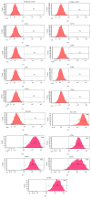
Histograms showing representative examples of DBMSCs isolated from passage three and stained with cell surface markers. DBMSCs were negative for the hematopoietic markers (CD14, CD19, and CD45), endothelial marker CD31, costimulatory molecules (CD40, CD80, CD83, and CD86), and HLA-DR. DBMSCs were positive for HLA-ABC, CD44, CD90, CD105, CD146, and CD166. Unstained and isotype controls were used ((a) and (b)). DBMSCs from passage three of 20 placentae were analyzed.
Figure 3
Figure 3
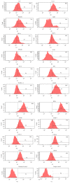
Histograms showing representative examples of DBMSCs isolated from passage three and stained with intracellular and cell surface markers. DBMSCs were positive for IL-6, IL-12, HMGB1, TLR-9, TNF-α, MCP-1, IFNAR1, IL5R, IL-10, IDO, TGFβ1,2,3, CD273, CD274, B7H3, B7H4, MMP-7, CXCL1, CXCL4, CXCL9, and CD130. Unstained and isotype controls were used ((u) and (v)). DBMSCs from passage three to passage five from five individual placentae were analyzed.
Figure 4
Figure 4
The effect of different cytokines on the proliferation of DBMSCs (a) and the effect of different chemotactic factors (HGF, SDF, MCP-1, and PDGF) on the migration of DBMSCs (b). The proliferation of DBMSCs was significantly increased in the presence of HGF, IL-6, IL-4, Rantes, and IL-17A ( P < 0.05). The chemotactic factors significantly ( P < 0.05) stimulated the migration of DBMSCs in transwell migration plates. DBMSCs at passage three from five individual placentae were analyzed. Bars represent standard errors.
Figure 5
Figure 5
Flow cytometric analysis of the expression of interferon gamma (IFN-γ), transforming growth factor-β (TGF-β), and intercellular adhesion molecule-1 (ICAM-1) by human decidua basalis mesenchymal stem cells (DBMSCs). The expression profiles of five individual experiments demonstrating that the expression of IFN-γ, TGF-β1, and ICAM-1 by DBMSCs significantly changed from passage 3 to passage 5. The levels of expression are presented as mean percentage of positive cells as determined by flow cytometry. Experiments were conducted in triplicate using the indicated DBMSC passages. Five independent placentae were used to prepare DBMSCs. P < 0.05. Bars represent standard errors.

References

    1. Friedenstein A. J., Chailakhjan R. K., Lalykina K. S. The development of fibroblast colonies in monolayer cultures of guinea-pig bone marrow and spleen cells. Cell and Tissue Kinetics. 1970;3(4):393–403. - PubMed
    1. Ding D.-C., Shyu W.-C., Lin S.-Z. Mesenchymal stem cells. Cell Transplantation. 2011;20(1):5–14. doi: 10.3727/096368910x. - DOI - PubMed
    1. Sethe S., Scutt A., Stolzing A. Aging of mesenchymal stem cells. Ageing Research Reviews. 2006;5(1):91–116. doi: 10.1016/j.arr.2005.10.001. - DOI - PubMed
    1. Tanaka M., Miyajima A. Identification and isolation of adult liver stem/progenitor cells. Methods in Molecular Biology. 2012;826:25–32. doi: 10.1007/978-1-61779-468-1_3. - DOI - PubMed
    1. Sakaguchi Y., Sekiya I., Yagishita K., Muneta T. Comparison of human stem cells derived from various mesenchymal tissues: superiority of synovium as a cell source. Arthritis and Rheumatism. 2005;52(8):2521–2529. doi: 10.1002/art.21212. - DOI - PubMed