. 2016 Mar 9:14:135-53.
doi: 10.1016/j.csbj.2016.02.005. eCollection 2016.

A guide to the identification of metabolites in NMR-based metabonomics/metabolomics experiments

Affiliations

A guide to the identification of metabolites in NMR-based metabonomics/metabolomics experiments

Anthony C Dona et al. Comput Struct Biotechnol J. .

Abstract

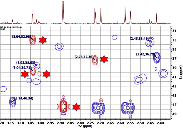
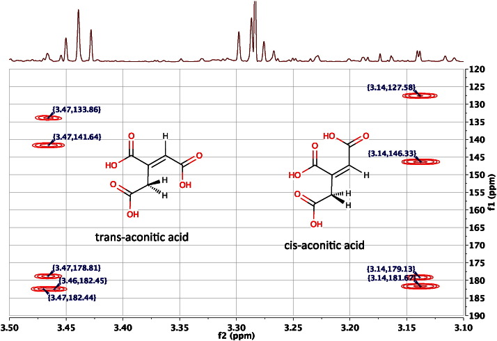
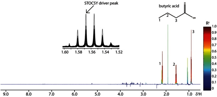
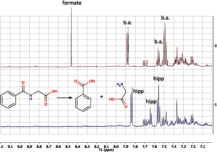
Metabonomics/metabolomics is an important science for the understanding of biological systems and the prediction of their behaviour, through the profiling of metabolites. Two technologies are routinely used in order to analyse metabolite profiles in biological fluids: nuclear magnetic resonance (NMR) spectroscopy and mass spectrometry (MS), the latter typically with hyphenation to a chromatography system such as liquid chromatography (LC), in a configuration known as LC-MS. With both NMR and MS-based detection technologies, the identification of the metabolites in the biological sample remains a significant obstacle and bottleneck. This article provides guidance on methods for metabolite identification in biological fluids using NMR spectroscopy, and is illustrated with examples from recent studies on mice.

Keywords: Metabolite identification; Metabolomics; Metabonomics; Molecular structure; Nuclear magnetic resonance (NMR) spectroscopy.


Figures

Fig. 1:
Fig. 1
an expansion of the 600 MHz 1H NMR spectrum of the urine of four, 30-week-old, male C57BL/6 mice, in the region of the doublet signals of citrate at ca 2.70 and 2.56 ppm. Even though the urine is buffered to pH 7.4, there are differences in the chemical shifts of the citrate signals between the four urine samples and noticeable differences also in half bandwidth, with the signals of mouse 1 (bottom spectrum) being especially broadened. The ‘roofing’ of the doublet citrate signals towards one another is illustrated by the arrows above the citrate resonances of mouse 4. See Section 2.6 on 2nd order effects.
Fig. 2
Fig. 2
An expansion of the 600 MHz 1H NMR spectrum of the urine of a male, 15-week-old, C57BL/6 mouse, in the region of the signal from the CH2–3 methylene protons of N-butyrylglycine at ca 1.62 ppm (dots). This pseudo-sextet signal is actually a triplet of quartets with the two 3JH,H couplings being almost equal in magnitude (7.4 and 7.5 Hz) resulting in overlap of many of the lines. The molecular structure of the metabolite is superimposed.
Fig. 3
Fig. 3
An expansion of the 600 MHz 1H NMR spectrum of the urine of pooled, male C57BL/6 mice at 15 weeks of age, in the region of the signal from the olefinic proton of cis-aconitic acid at ca 5.74 ppm. The signal is a triplet due to a long-range, 4-bond, 4JH,H coupling of ca 1.4 Hz to the two equivalent methylene hydrogens. The 1:2:1 nature of the triplet is clear, even though it is superimposed upon the very broad signal from urea at ca 5.80 ppm.
Fig. 4
Fig. 4
An expansion of the 600 MHz 1H NMR spectrum of the pooled urine of male C57BL/6 mice at 15 weeks of age, in the region of the signal from the methyl protons of choline (structure superimposed) at ca 3.20 ppm. The signal is a 1:1:1 triplet (dots) due to a 2-bond coupling of ca 0.6 Hz to the 14N nucleus. Interestingly, the well-resolved doublet at ca 3.13 ppm is due to the methylene protons of cis-aconitic acid with 4JH,H coupling of ca 1.4 Hz (see also Fig. 3). The spectrum has been zero filled to 131,072 points and resolution enhanced by Gaussian multiplication, prior to Fourier transformation.
Fig. 5
Fig. 5
Two versions of the 600 MHz 1H NMR spectrum of an authentic sample of the metabolite para-cresol sulphate in deuterated phosphate buffer at pH 7.4, in the region of the signals from the aromatic hydrogens: 1) with a standard 0.3 Hz line-broadening and 2) resolution-enhanced using a Lorentzian to Gaussian transformation. The signal of the H2, H6 protons appears as a complex, second-order multiplet at ca 7.22 ppm, instead of a first-order doublet. The signal of the H3, H5 protons at ca 7.29 ppm displays additional complexity due to coupling to the methyl protons via a 4-bond coupling, in addition to the extra lines, clearly visible in this second-order system.
Fig. 6
Fig. 6
1) The 600 MHz 1H NMR spectrum of the urine of a 30-week-old, male, flavin mono-oxygenase 5 (FMO5) knockout mouse in the region of the aromatic signals from hippuric acid (structure inset). The spectrum is resolution enhanced by Gaussian multiplication. 2) A spin simulation of the aromatic signals from hippuric acid using the MNova spin simulation function. A good approximation to the complex, second-order signals was obtained. The complexity of the two ortho and two meta hydrogen signals is due to the fact that whilst these hydrogens are chemically equivalent (within each pair), they are magnetically non-equivalent and are part of a five hydrogen AA’BB’M spin system (see Section 2.6). Signals from 3-indoxyl sulphate and other metabolites are present in the real spectrum (1).
Fig. 7
Fig. 7
The low frequency region of the 600 MHz 2D 1H J-resolved NMR spectrum of the urine of a male, 30-week-old, FMO5 knockout mouse displayed as a contour plot underneath the corresponding 1D 1H NMR spectrum. The overlapping signals from the triplet methyl group of N-butyrylglycine (0.926 ppm, three blue circles and downward arrows) and the doublet methyl group of isovaleric acid (0.916 ppm, two red squares and upward arrows) are completely resolved in the 2D JRES NMR spectrum. The spectrum is tilted by 450, so that all the signals of each multiplet appear at the same chemical shift, and it is symmetrised.
Fig. 8
Fig. 8
An expansion of the 600 MHz 2D 1H J-resolved NMR spectrum of the urine of a male, 30-week-old, FMO5 knockout mouse in the region of the AB resonances from citric acid at ca 2.70 and ca 2.56 ppm (four dots in 1D spectrum), displayed as a contour plot underneath the corresponding 1D 1H NMR spectrum. The spectrum is tilted by 450, so that all the signals of each multiplet appear at the same chemical shift, and symmetrised. The signals labelled with stars, appearing at ca 2.63 ppm, exactly in between the shifts of the two citrate signals are 2nd order effects caused by the mixing of transitions between the A and B spins by the 1800 pulse, in the presence of strong coupling. As is clear from the 1D 1H NMR spectrum, there are no real signals at 2.63 ppm!
Fig. 9
Fig. 9
An expansion of the 600 MHz 2D 1H J-resolved NMR spectrum of the urine of a male, 30-week-old, FMO5 knockout mouse in the region of the resonances from the C3 methylene hydrogens of 2S-hydroxyglutaric acid, displayed as a contour plot underneath the corresponding 1D 1H NMR spectrum. The spectrum is tilted by 450, so that all the signals of each multiplet appear at the same chemical shift, and symmetrised. The peak picking allows a simple analysis of three of the four couplings that these hydrogens possess as 4.2, 6.3 and 10.5 Hz (2.003 ppm) and 5.5, 7.6 and 10.3 Hz (1.845 ppm). Note that these multiplets are invisible in the 1D 1H NMR spectrum.
Fig. 10
Fig. 10
An expansion of the 600 MHz 2D 1H COSY NMR spectrum of the pooled urine of two male, 60-week-old, FMO5 knockout mice in the region of the broad singlet methyl resonances from trigonelline at ca 4.435 and 1-methylnicotinamide ca 4.475 ppm, displayed as a contour plot underneath the corresponding resolution-enhanced 1D 1H NMR spectrum. Trigonelline displays cross-peaks due to long-range, 4-bond coupling from the methyl protons to the H2 (9.111) and H6 (8.820) protons ortho to the pyridinium nitrogen. 1-methylnicotinamide displays the same cross-peaks to H2 (9.259) and H6 (8.951), but in addition, displays a clear and remarkable cross-peak via six-bond coupling to H4 (8.883). The ability to connect the methyl shift with the pyridinium proton shifts in this way can assist metabolite identification enormously.
Fig. 11
Fig. 11
An expansion of the 600 MHz 2D 1H COSY NMR spectrum of the urine of a male, 30-week-old, FMO5 knockout mouse highlighting with 7 arrows the cross-peak from the C4H proton of ketoleucine (structure inset) at 2.098 to the equivalent C5 and C6 methyl groups at 0.941 ppm. The signals from ketoleucine at 2.098 are not visible either in the 1D 1H NMR spectrum (top), or in the 2D 1H J-resolved NMR spectrum of the same sample (see Fig. 9) but the identification is confirmed from this high-resolution COSY spectrum. The seven cross peaks marked are the most intense peaks of the 9-line, pseudo-nonet, triplet of septets, the two outside lines of which are too weak to observe. See text for details.
Fig. 12
Fig. 12
An expansion of the 600 MHz 2D 1H TOCSY NMR spectrum of the urine of a 30-week-old male C57BL/6 mouse. The cross peaks marked originate from the alkyl chain connectivities of N-butyrylglycine, from the terminal methyl group (C4). The cross peak marked at 0.926, 1.617 ppm represents a direct correlation from the C4 methyl protons to the adjacent C3 methylene group, equivalent to the cross-peak that would be observed in a 2D 1H COSY experiment. Additional metabolite identification information is provided in this TOCSY experiment however, with the cross-peak at 0.926, 2.279 ppm establishing a connection between the C4 methyl protons and the C2 methylene group, even though there is no observable coupling between them.
Fig. 13
Fig. 13
An expansion of the 600 MHz, multiplicity-edited, 2D 13C, 1H HSQC NMR spectrum of the pooled urine of 60-week-old, male, FMO5 knockout mice , displayed as a contour plot underneath the corresponding resolution-enhanced 1D 1H NMR spectrum. In this phase-sensitive plot, positive peaks are represented by red contours (asterisked) and negative peaks by blue contours (no asterisks). See text for further explanation.
Fig. 14
Fig. 14
An expansion of the 600 MHz 2D 13C, 1H HMBC NMR spectrum of the pooled urine of male, 60-week-old, FMO5 knockout mice , displayed as a contour plot underneath the corresponding resolution-enhanced 1D 1H NMR spectrum in the region of the signals from the methylene protons of cis-aconitic acid (3.14) and trans-aconitic acid (3.47 ppm). The methylene protons display all four possible 2- and 3-bond hydrogen-to-carbon connectivities, to both adjacent carboxylic acid carbons (178.8, 182.4 ppm, trans- and 179.1 and 181.7 ppm, cis-isomer) plus connections to the quaternary and protonated olefinic carbons at 141.6 and 133.9 (trans-) and 146.3 and 127.6 ppm (cis-isomer), respectively, thus establishing connectivities between the two regions of protonated carbon structure isolated from each other by the quaternary olefinic carbon.
Fig. 15
Fig. 15
NMR plot following a STOCSY analysis on a set of faecal water 1H NMR spectra. The selected driver peak at 1.57 ppm was used to calculate the correlation matrix which reveals correlations ranging from 0 (low) to 1 (high). Two other resonances were revealed to have a positive correlation of 1, suggesting that they arise from the same molecule that was later identified as butyric acid.
Fig. 16
Fig. 16
An expansion of the 600 MHz 1H NMR spectra of the urine of a male, 30-week-old, FMO5 knockout mouse : 1) before bacterial fermentation and 2) after bacterial fermentation after leaving the sample at ambient temperature for several days. The bacterial fermentation caused many metabolic transformations including that of hippuric acid (hipp) to benzoic acid (b.a.) and glycine (3.57 ppm, not shown) and the formation of formate. The lower spectrum 1) prior to fermentation shows many signals including those from the ortho (7.84), para (7.64) and meta (7.56) protons of hippuric acid, whereas post-fermentation, spectrum 2) at top, shows corresponding signals from the ortho (7.88), para (7.56) and meta (7.49 ppm) protons of benzoic acid.
Image 1
Image 3
Image 5

References

    1. Lindon J., Nicholson J., Holmes E., Everett J. Metabonomics: metabolic processes studied by NMR spectroscopy of biofluids. Concepts Magn Reson. 2000;12(5):289–320.
    1. Fiehn O. Metabolomics—the link between genotypes and phenotypes. Plant Mol Biol. 2002;48(1–2):155–171. - PubMed
    1. Lindon J.C., Nicholson J.K., Holmes E. Elsevier; Amsterdam, Oxford: 2007. The Handbook of Metabonomics and Metabolomics.
    1. Clayton T., Lindon J., Cloarec O., et al. Pharmaco-metabonomic phenotyping and personalized drug treatment. Nature. 2006;440(7087):1073–1077. - PubMed
    1. Clayton T.A., Baker D., Lindon J.C., Everett J.R., Nicholson J.K. Pharmacometabonomic identification of a significant host–microbiome metabolic interaction affecting human drug metabolism. Proc Natl Acad Sci U S A. 2009;106(34):14728–14733. - PMC - PubMed