

Ultrasound of paediatric appendicitis and its secondary sonographic signs: providing a more meaningful finding
- PMID: 27087976
- PMCID: PMC4775827
- DOI: 10.1002/jmrs.154
Ultrasound of paediatric appendicitis and its secondary sonographic signs: providing a more meaningful finding
Abstract
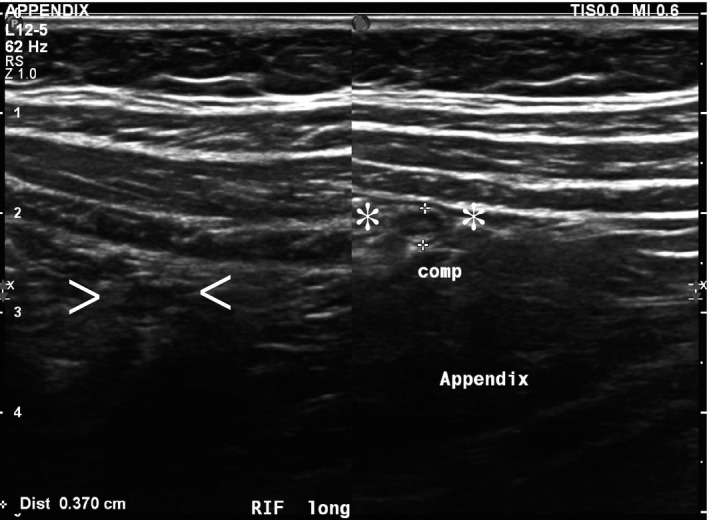
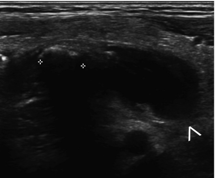
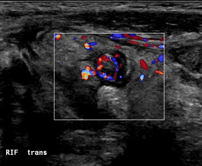
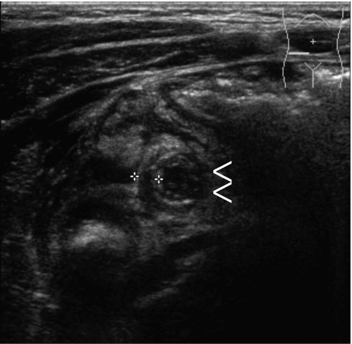
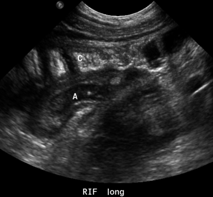
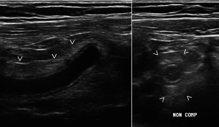
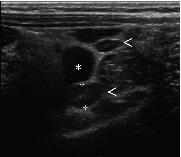
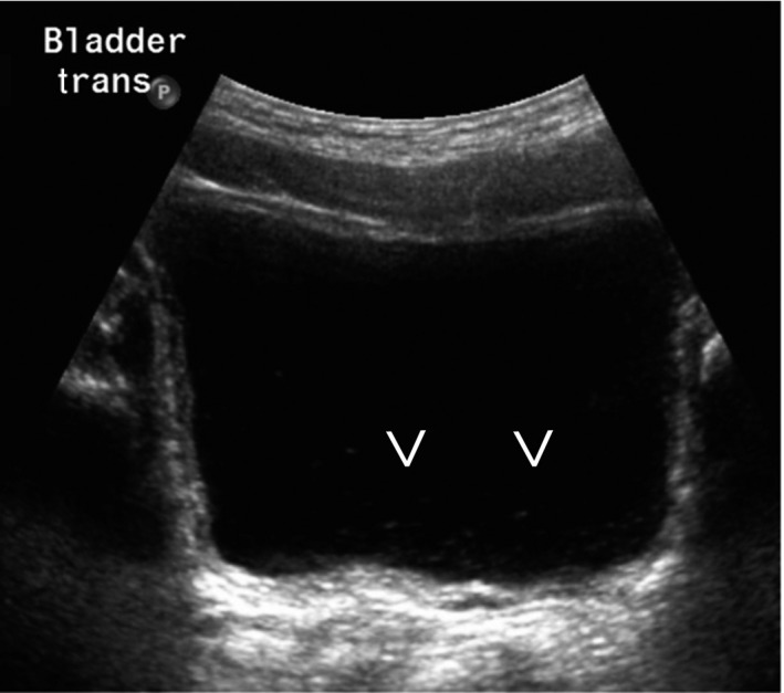
Sonography is an important clinical tool in diagnosing appendicitis in children as it can obviate both exposure to potentially harmful ionising radiation from computed tomography scans and the need for unnecessary appendicectomies. This review examines the diagnostic accuracy of ultrasound in the identification of acute appendicitis, with a particular focus on the the utility of secondary sonographic signs as an adjunct or corollary to traditionally examined criteria. These secondary signs can be important in cases where the appendix cannot be identified with ultrasound and a more meaningful finding may be made by incorporating the presence or absence of secondary sonographic signs. There is evidence that integrating these secondary signs into the final ultrasound diagnosis can improve the utility of ultrasound in cases where appendicitis is expected, though there remains some conjecture about whether they play a more important role in negative or positive prediction in the absence of an identifiable appendix.
Keywords: Appendicitis; child; paediatrics; ultrasonography.
Figures
References
-
- Australian Institute of Health and Welfare . Admitted Patient Care 2013–14: Australian Hospital Statistics. Health services series no. 60. Cat. no. HSE 156. AIHW, Canberra, Australia, 2015.
-
- Sivit CJ, Applegate KE, Stallion A, et al. Imaging evaluation of suspected appendicitis in a pediatric population. Am J Roentgenol 2000; 175: 977–80. - PubMed
-
- Wiersma F, Toorenvliet BRB, Bloem JJL, Allema JH, Holscher HC. US examination of the appendix in children with suspected appendicitis: the additional value of secondary signs. Eur Radiol 2009; 19: 455–61. - PubMed
-
- Gracey D, McClure MJ. The impact of ultrasound in suspected acute appendicitis. Clin Radiol 2007; 62: 573–8. - PubMed
-
- Baldisserotto M, Marchiori E. Accuracy of noncompressive sonography of children with appendicitis according to the potential positions of the appendix. AJR Am J Roentgenol 2000; 175: 1387–92. - PubMed
Publication types
MeSH terms
LinkOut - more resources
Full Text Sources
Other Literature Sources
Medical
