

Review
doi: 10.1038/mtm.2016.14.
eCollection 2016.
From selection hits to clinical leads: progress in aptamer discovery
Affiliations
- PMID: 27088106
- PMCID: PMC4822646
- DOI: 10.1038/mtm.2016.14
Item in Clipboard
Review
From selection hits to clinical leads: progress in aptamer discovery
Mol Ther Methods Clin Dev.
.
Abstract
Aptamers were discovered more than 25 years ago, yet only one has been approved by the US Food and Drug Administration to date. With some noteworthy advances in their chemical design and the enzymes we use to make them, aptamers and aptamer-based therapeutics have seen a resurgence in interest. New aptamer drugs are being approved for clinical evaluation, and it is certain that we will see increasingly more aptamers and aptamer-like drugs in the future. In this review, we will discuss the production of aptamers with an emphasis on the advances and modifications that enabled early aptamers to succeed in clinical trials as well as those that are likely to be important for future generations of these drugs.
Figures
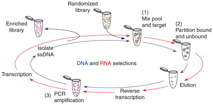
A schematic of the selection process for DNA and RNA aptamer libraries. Starting with randomized library incubated with the target (1), bound species are partitioned and stringently washed (2), followed by elution of desired species. For RNA selections, recovered material must be reverse transcribed, followed by polymerase chain reaction (PCR) amplification (3) and transcription back into RNA to generate the library for the next round. DNA selections however, are ready for PCR amplification after elution (3), but afterwards must be separated from the complement strand before the resulting ssDNA pool can be used for the next round.
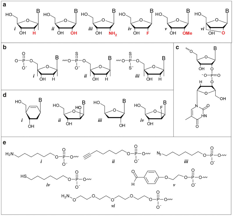
Modifications utilized to enhance the in vivo stability of aptamers. (a) 2’-modifications can easily be incorporated into aptamers during chemical synthesis and include i. 2’H, ii. 2’OH, iii. 2’ NH2, iv. 2’F, v. 2’OMe and vi. locked nucleic acids. (b) Increased stability can also be garnered though thiolation of the phosphate backbone. Structures shown (from left to right) are for the i. natural phosphodiester, ii. the thiolated phosothioate and iii. phosphorodithioate. (c) 3’ inverted deoxythymidine residue. (d) Non-ribose backbones, which can be incorporated using novel DNA polymerases for the basis for xeno nucleic acids, include i. cyclohexenyl, ii. arabino, iii. α-L-threofuranosy, and iv. 2′-fluoroarabino nucleic acids. (e) Examples of some commercially available functional groups that can be readily attached to the 5’-end during solid phase synthesis and used to facilitate downstream conjugation include i. amine, ii. alkyne, iii. azide, iv. thiol, v. aldehyde and vi. aminooxy.
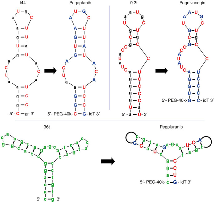
Only the three aptamers have reached Phase III clinical trials to date. Minimized lead molecules as produced from the selection shown next to their counterpart fully stabilized clinical progeny. Lowercase black and green denotes 2’OH and 2’H respectively, uppercase red and blue denotes 2’F and 2’OMe respectively. “idT” represents an inverted deoxythimidine, also known as 3’-3’ dT. PEG-40k represents an amine linked to 40 kDa polyethylene glycol. Black loops on the arms of pegpluranib represents an 18-atom hexaethylene glycol spacer, which replaced the 3 nucleotide loops found in the parent molecule.
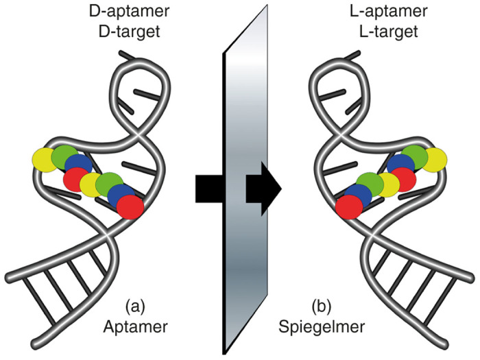
Mirror image aptamers, Spiegelmers, are composed of non-natural L-ribose nucleotides. The molecules are initially selected from natural D-ribose aptamer libraries against a non-natural target, for example a D-peptide (a). Once optimized as a D-aptamer the mirror image L-aptamer (Spiegelmer) is synthesized chemically and intrinsically binds to the natural L-target, such as a naturally occurring protein (b).
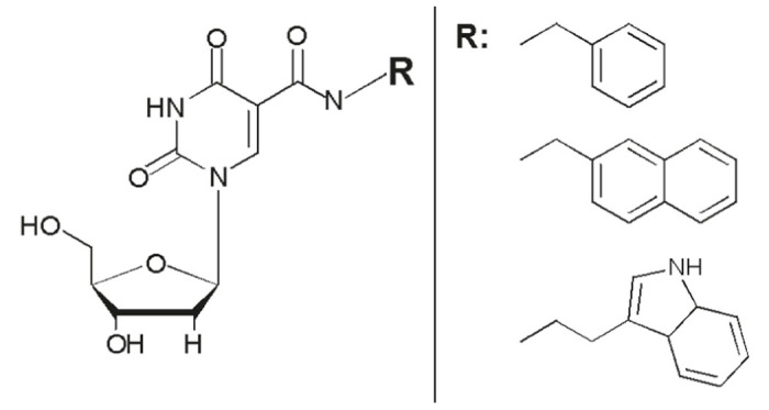
Modified deoxyuridine (dU) residues are at the core of the novel molecules developed by Somalogic. A variety of chemical moieties are attached to the 5-position of dU via a carboxyamide linkage (left). A variety of different modifications (R) have been employed for the selection of SOMAmers including benzyl, napthyl, and indole (right).
References
-
- Tuerk, C and Gold, L (1990). Systematic evolution of ligands by exponential enrichment: RNA ligands to bacteriophage T4 DNA polymerase. Science 249: 505–510. - PubMed
-
- Ellington, AD and Szostak, JW (1990). In vitro selection of RNA molecules that bind specific ligands. Nature 346: 818–822. - PubMed
-
- Hall, B, Arshad, S, Seo, K, Bowman, C, Corley, M, Jhaveri, SD et al. (2009). In Vitro Selection of RNA Aptamers to a Protein Target by Filter Immobilization. John Wiley & SonsHoboken. pp. 24.3.1–24.3.27. - PubMed
-
- Yan, A and Levy, M (2014). Cell internalization SELEX: in vitro selection for molecules that internalize into cells. Methods Mol Biol 1103: 241–265. - PubMed
Publication types
LinkOut - more resources
Full Text Sources
Other Literature Sources
