

Voltage-gated calcium channel autoimmune cerebellar degeneration: Case and study of cytotoxicity
- PMID: 27088118
- PMCID: PMC4821674
- DOI: 10.1212/NXI.0000000000000222
Voltage-gated calcium channel autoimmune cerebellar degeneration: Case and study of cytotoxicity
Abstract
Objectives: To describe response to treatment in a patient with autoantibodies against voltage-gated calcium channels (VGCCs) who presented with autoimmune cerebellar degeneration and subsequently developed Lambert-Eaton myasthenic syndrome (LEMS), and to study the effect of the patient's autoantibodies on Purkinje cells in rat cerebellar slice cultures.
Methods: Case report and study of rat cerebellar slice cultures incubated with patient VGCC autoantibodies.
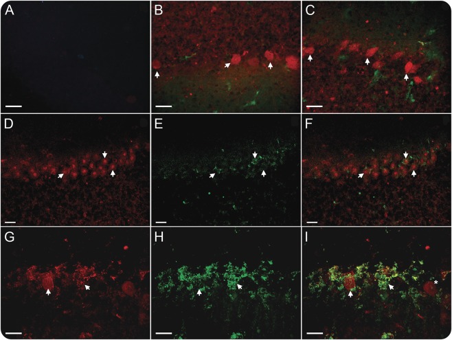
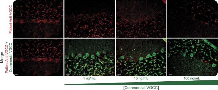
Results: A 53-year-old man developed progressive incoordination with ataxic speech. Laboratory evaluation revealed VGCC autoantibodies without other antineuronal autoantibodies. Whole-body PET scans 6 and 12 months after presentation detected no malignancy. The patient improved significantly with IV immunoglobulin G (IgG), prednisone, and mycophenolate mofetil, but worsened after IV IgG was halted secondary to aseptic meningitis. He subsequently developed weakness with electrodiagnostic evidence of LEMS. The patient's IgG bound to Purkinje cells in rat cerebellar slice cultures, followed by neuronal death. Reactivity of the patient's autoantibodies with VGCCs was confirmed by blocking studies with defined VGCC antibodies.
Conclusions: Autoimmune cerebellar degeneration associated with VGCC autoantibodies may precede onset of LEMS and may improve with immunosuppressive treatment. Binding of anti-VGCC antibodies to Purkinje cells in cerebellar slice cultures may be followed by cell death. Patients with anti-VGCC autoantibodies may be at risk of irreversible neurologic injury over time, and treatment should be initiated early.
Figures
Similar articles
-
Paraneoplastic cerebellar degeneration. III. Cerebellar degeneration, cancer, and the Lambert-Eaton myasthenic syndrome.Neurology. 1992 Oct;42(10):1944-50. doi: 10.1212/wnl.42.10.1944. Neurology. 1992. PMID: 1407577 Review.
-
Pathogenic autoantibodies in the lambert-eaton myasthenic syndrome.Ann N Y Acad Sci. 2003 Sep;998:187-95. doi: 10.1196/annals.1254.019. Ann N Y Acad Sci. 2003. PMID: 14592874
-
Reduction of P/Q-type calcium channels in the postmortem cerebellum of paraneoplastic cerebellar degeneration with Lambert-Eaton myasthenic syndrome.Ann Neurol. 2003 Jan;53(1):21-8. doi: 10.1002/ana.10392. Ann Neurol. 2003. PMID: 12509844
-
[Paraneoplastic cerebellar degeneration and Lambert-Eaton myasthenic syndrome associated with anti P/Q-type voltage-gated calcium channel antibody in a patient with primary double lung cancer].Brain Nerve. 2009 Sep;61(9):1083-7. Brain Nerve. 2009. PMID: 19803409 Japanese.
-
[Autoantibody against the presynaptic P/Q-type voltage-gated calcium channel in Lambert-Eaton myasthenic syndrome].Brain Nerve. 2013 Apr;65(4):441-8. Brain Nerve. 2013. PMID: 23568992 Review. Japanese.
Cited by
-
Paraneoplastic and Other Autoimmune Encephalitides: Antineuronal Antibodies, T Lymphocytes, and Questions of Pathogenesis.Front Neurol. 2022 Jan 17;12:744653. doi: 10.3389/fneur.2021.744653. eCollection 2021. Front Neurol. 2022. PMID: 35111121 Free PMC article. Review.
-
Neuroimmunogastroenterology: At the Interface of Neuroimmunology and Gastroenterology.Front Neurol. 2020 Jul 31;11:787. doi: 10.3389/fneur.2020.00787. eCollection 2020. Front Neurol. 2020. PMID: 32849234 Free PMC article. Review.
-
Late onset cerebellar ataxia syndrome after non-paraneoplastic Lambert-Eaton myasthenic syndrome: a case study.BMC Neurol. 2025 Jan 2;25(1):2. doi: 10.1186/s12883-024-03983-8. BMC Neurol. 2025. PMID: 39748289 Free PMC article.
-
Movement disorders with neuronal antibodies: syndromic approach, genetic parallels and pathophysiology.Brain. 2018 Jan 1;141(1):13-36. doi: 10.1093/brain/awx189. Brain. 2018. PMID: 29053777 Free PMC article. Review.
-
A Breakdown of Immune Tolerance in the Cerebellum.Brain Sci. 2022 Feb 28;12(3):328. doi: 10.3390/brainsci12030328. Brain Sci. 2022. PMID: 35326284 Free PMC article. Review.
References
-
- Greenlee JE. Treatment of paraneoplastic neurologic disorders. Curr Treat Options Neurol 2010;12:212–230. - PubMed
-
- Blumenfeld AM, Recht LD, Chad DA, DeGirolami U, Griffin T, Jaeckle KA. Coexistence of Lambert-Eaton myasthenic syndrome and subacute cerebellar degeneration: differential effects of treatment. Neurology 1991;41:1682–1685. - PubMed
-
- Clouston PD, Saper CB, Arbizu T, et al. Paraneoplastic cerebellar degeneration: III: cerebellar degeneration, cancer, and the Lambert-Eaton myasthenic syndrome. Neurology 1992;42:1944–1950. - PubMed
-
- Goldstein JM, Waxman SG, Vollmer TL, Lang B, Johnston I, Newsom-Davis J. Subacute cerebellar degeneration and Lambert-Eaton myasthenic syndrome associated with antibodies to voltage-gated calcium channels: differential effect of immunosuppressive therapy on central and peripheral defects. J Neurol Neurosurg Psychiatry 1994;57:1138–1139. - PMC - PubMed
LinkOut - more resources
Full Text Sources
Other Literature Sources