. 2015 Dec;1(4):252-265.
doi: 10.1016/j.trecan.2015.10.009.

Glioblastoma: Defining Tumor Niches

Affiliations

Glioblastoma: Defining Tumor Niches

Dolores Hambardzumyan et al. Trends Cancer. 2015 Dec.

Abstract

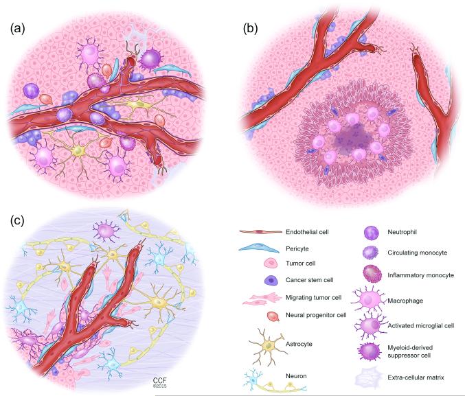
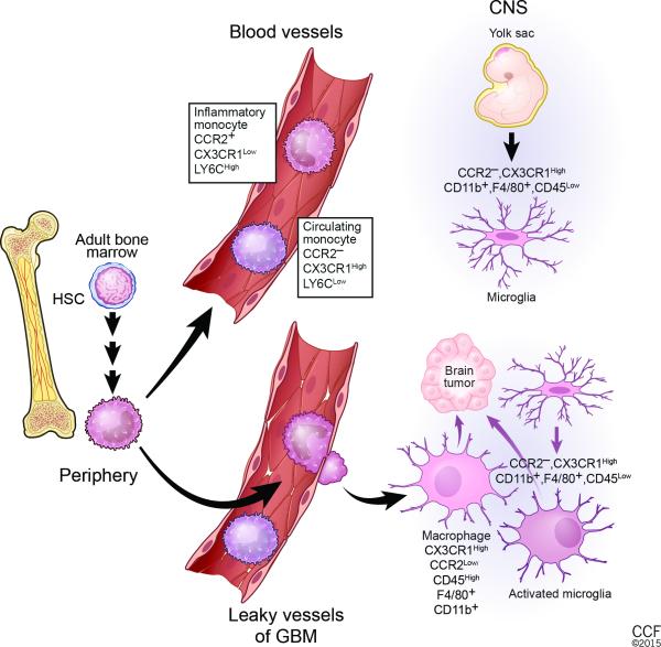
Glioblastomas (GBM) are one of the most recalcitrant brain tumors because of their aggressive invasive growth and resistance to therapy. They are highly heterogeneous malignancies at both the molecular and histological levels. Specific histological hallmarks including pseudopalisading necrosis and microvascular proliferation distinguish GBM from lower-grade gliomas, and make GBM one of the most hypoxic as well as angiogenic tumors. These microanatomical compartments present specific niches within the tumor microenvironment that regulate metabolic needs, immune surveillance, survival, invasion as well as cancer stem cell maintenance. Here we review features and functions of the distinct GBM niches, detail the different cell constituents and the functional status of the vasculature, and discuss prospects of therapeutically targeting GBM niche constituents.

Keywords: GBM; astrocytes; cancer stem cells; macrophages; microglial cells; therapy; tumor microenvironment; tumor niches; tumor vasculature.


Figures

Figure 1
Figure 1. Illustrations of the different Glioblastoma (GBM) niches
(a) The perivascular GBM niche (PVN). Non-neoplastic cell and glioma cell interactions create a specialized vascular niche, which provides a supportive environment for cancer stem cell (CSC) growth, maintenance, and survival. The PVN niche is a multi-cellular structure composed of non-neoplastic cells, including endothelial cells, pericytes, macrophages, neutrophils, myeloid-derived suppressor cells (MDSCs), reactive astrocytes, and infiltrating neural progenitor cells (NPCs), as well as neoplastic cells including tumor bulk (TB) and CSCs. Macrophages and other non-neoplastic cells are recruited to the tumor by TB and CSCs. These recruited and reprogrammed non-neoplastic cells secrete soluble factors that expand the TB and CSCs and establish a supportive niche for CSCs. In addition, interactions between reactive astrocytes and TB and CSCs can lead to further tumor growth. Pericytes interact with CSCs and TB to promote tumor growth and to contribute to leakiness of the blood brain barrier (BBB). In contrast to normal brain microvessels, both astrocyte and pericyte coverage is incomplete in GBM vessels. Lastly, each cellular component has the capacity to change the extracellular matrix composition that impacts glioma proliferation, survival, and expansion. (b) The hypoxic GBM niche. The pseudopalisading areas with a necrotic core are hallmarks of GBM and create the hypoxic niche for CSCs. Hypoxia through induction of hypoxia inducible factor 1 alpha (HIF-1α) and of hypoxia inducible factor 2 alpha (HIF-2α) promotes the expansion of cancer stem cells (CSCs) and recruits innate immune cells including macrophages. Hypoxia generally occurs when tumor growth exceeds neovascularization. (c) The invasive GBM niche. Glioma cells migrate along blood vessels at the invasive edge that is defined as perivascular invasion. Major cell types that constitute the microenvironment at the invasive edge of GBM include endothelial cells, pericytes, activated microglia, reactive astrocytes and neurons. Invading glioma cells make surgical resection incomplete and are partially responsible for tumor recurrence.
Fig. 2
Fig. 2. Cellular origin of GBM-associated microglia and monocytes
Under steady-state conditions, microglia and monocytes reside in separate locations. In adult life, monocytes are generated from hematopoietic stem cells (HSCs) via a series of intermediate progenitors. Ly6CLow, CCR2, CX3CR1High circulating monocytes and Ly6CHigh, CCR2+, CX3CR1Low inflammatory monocytes are released into the blood circulation. Inflammatory monocytes are recruited to sites of pathological conditions like GBM. Once within the central nervous system (CNS), they can differentiate into tumor-associated macrophages and become phenotypically nearly indistinguishable from activated resident microglia. Microglia are resident macrophages in the brain that originate from yolk sac progenitors in the neuroepithelium beginning around embryonic day 8.5 in the mouse. In the adult brain, they are negative for CCR2 but express high levels of CX3CR1, CD11b, F4/80, and low levels of CD45. GBM cells induce local inflammation that compromises the integrity of the BBB and results in tumor infiltration of inflammatory monocytes and attracts brain-resident microglial cells to glioma margins.

References

    1. Stupp R, et al. Radiotherapy plus concomitant and adjuvant temozolomide for glioblastoma. N Engl J Med. 2005;352:987–996. - PubMed
    1. Brennan CW, et al. The somatic genomic landscape of glioblastoma. Cell. 2013;155:462–477. - PMC - PubMed
    1. Cancer Genome Atlas Research, N. Comprehensive genomic characterization defines human glioblastoma genes and core pathways. Nature. 2008;455:1061–1068. - PMC - PubMed
    1. Verhaak RG, et al. Integrated genomic analysis identifies clinically relevant subtypes of glioblastoma characterized by abnormalities in PDGFRA, IDH1, EGFR, and NF1. Cancer Cell. 2010;17:98–110. - PMC - PubMed
    1. Snuderl M, et al. Mosaic amplification of multiple receptor tyrosine kinase genes in glioblastoma. Cancer Cell. 2011;20:810–817. - PubMed