

Crystal structure of glycogen debranching enzyme and insights into its catalysis and disease-causing mutations
- PMID: 27088557
- PMCID: PMC4837477
- DOI: 10.1038/ncomms11229
Crystal structure of glycogen debranching enzyme and insights into its catalysis and disease-causing mutations
Abstract
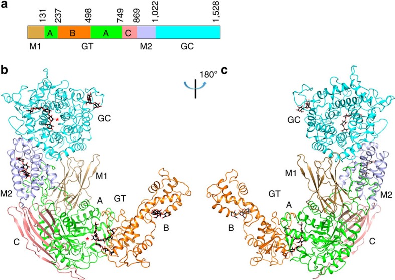
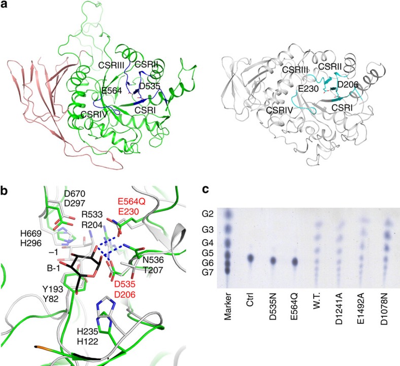
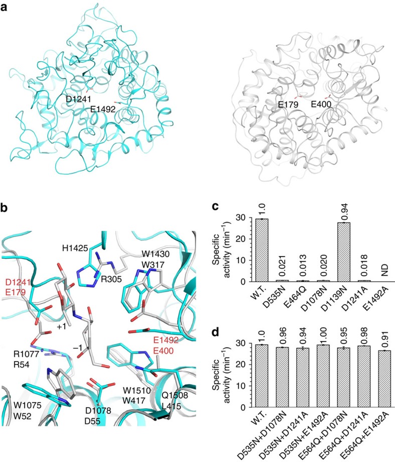
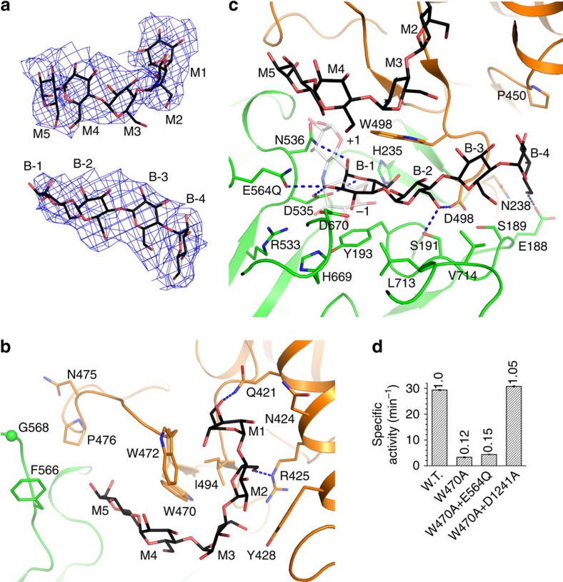
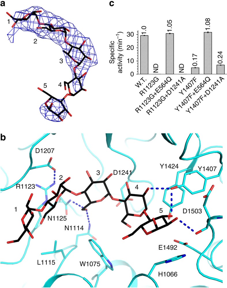
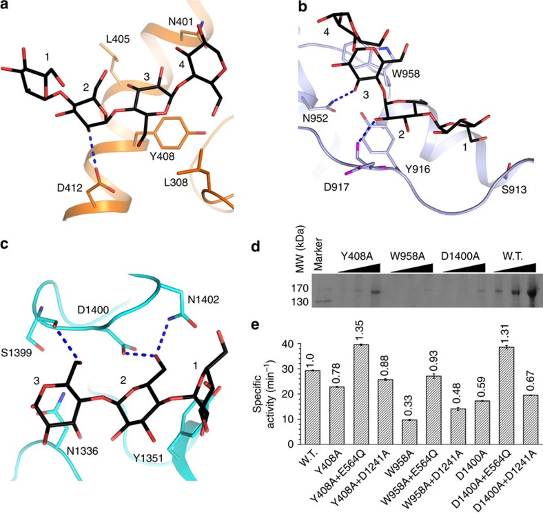
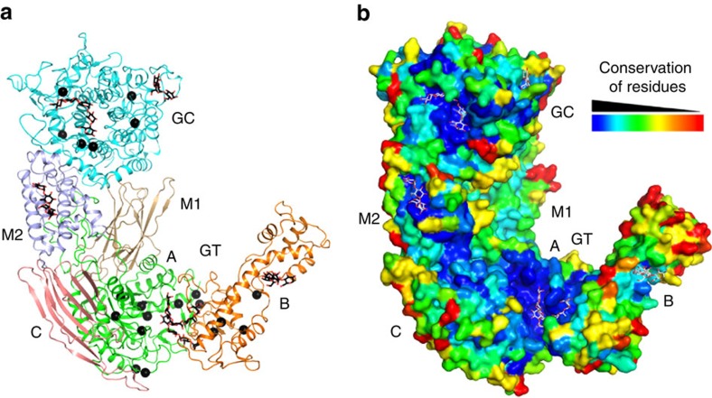
Glycogen is a branched glucose polymer and serves as an important energy store. Its debranching is a critical step in its mobilization. In animals and fungi, the 170 kDa glycogen debranching enzyme (GDE) catalyses this reaction. GDE deficiencies in humans are associated with severe diseases collectively termed glycogen storage disease type III (GSDIII). We report crystal structures of GDE and its complex with oligosaccharides, and structure-guided mutagenesis and biochemical studies to assess the structural observations. These studies reveal that distinct domains in GDE catalyse sequential reactions in glycogen debranching, the mechanism of their catalysis and highly specific substrate recognition. The unique tertiary structure of GDE provides additional contacts to glycogen besides its active sites, and our biochemical experiments indicate that they mediate its recruitment to glycogen and regulate its activity. Combining the understanding of the GDE catalysis and functional characterizations of its disease-causing mutations provides molecular insights into GSDIII.
Figures
Similar articles
-
Crystal structures of glycogen-debranching enzyme mutants in complex with oligosaccharides.Acta Crystallogr F Struct Biol Commun. 2021 Nov 1;77(Pt 11):420-426. doi: 10.1107/S2053230X21010918. Epub 2021 Oct 29. Acta Crystallogr F Struct Biol Commun. 2021. PMID: 34726181 Free PMC article.
-
Molecular architecture and catalytic mechanism of human glycogen debranching enzyme.Nat Commun. 2025 Jul 1;16(1):5962. doi: 10.1038/s41467-025-61077-6. Nat Commun. 2025. PMID: 40593796 Free PMC article.
-
Mutations in exon 3 of the glycogen debranching enzyme gene are associated with glycogen storage disease type III that is differentially expressed in liver and muscle.J Clin Invest. 1996 Jul 15;98(2):352-7. doi: 10.1172/JCI118799. J Clin Invest. 1996. PMID: 8755644 Free PMC article.
-
Molecular characterization of glycogen storage disease type III.Curr Mol Med. 2002 Mar;2(2):167-75. doi: 10.2174/1566524024605752. Curr Mol Med. 2002. PMID: 11949933 Review.
-
[Glycogen debranching enzyme deficiency (Forbes-Cori disease)].Ryoikibetsu Shokogun Shirizu. 2001;(36):20-2. Ryoikibetsu Shokogun Shirizu. 2001. PMID: 11596370 Review. Japanese. No abstract available.
Cited by
-
A functional mini-GDE transgene corrects impairment in models of glycogen storage disease type III.J Clin Invest. 2024 Jan 16;134(2):e172018. doi: 10.1172/JCI172018. J Clin Invest. 2024. PMID: 38015640 Free PMC article.
-
Multi-omics analysis to examine microbiota, host gene expression and metabolites in the intestine of black tiger shrimp (Penaeus monodon) with different growth performance.PeerJ. 2020 Aug 14;8:e9646. doi: 10.7717/peerj.9646. eCollection 2020. PeerJ. 2020. PMID: 32864208 Free PMC article.
-
Crystal structures of glycogen-debranching enzyme mutants in complex with oligosaccharides.Acta Crystallogr F Struct Biol Commun. 2021 Nov 1;77(Pt 11):420-426. doi: 10.1107/S2053230X21010918. Epub 2021 Oct 29. Acta Crystallogr F Struct Biol Commun. 2021. PMID: 34726181 Free PMC article.
-
Role of Metabolism in Bone Development and Homeostasis.Int J Mol Sci. 2020 Nov 26;21(23):8992. doi: 10.3390/ijms21238992. Int J Mol Sci. 2020. PMID: 33256181 Free PMC article. Review.
-
Glycogen storage diseases.Nat Rev Dis Primers. 2023 Sep 7;9(1):46. doi: 10.1038/s41572-023-00456-z. Nat Rev Dis Primers. 2023. PMID: 37679331 Review.
References
-
- Roach P. J. Glycogen and its metabolism. Curr. Mol. Med. 2, 101–120 (2002). - PubMed
-
- Dagli A., Sentner C. P. & Weinstein D. A. Glycogen Storage Disease Type III in (eds Pagon, R. A., Adam, M. P., Ardinger, H. H). Available at: http://www.ncbi.nlm.nih.gov/books/NBK26372/ (2012).
-
- Nelson T. E., Kolb E. & Larner J. Purification and properties of rabbit muscle amylo-1,6-glucosidase-oligo-1,4-1,4-transferase. Biochemistry 8, 1419–1428 (1969). - PubMed
Publication types
MeSH terms
Substances
LinkOut - more resources
Full Text Sources
Other Literature Sources
