

Prenatal Diagnosis of Lissencephaly Type 2 using Three-dimensional Ultrasound and Fetal MRI: Case Report and Review of the Literature
- PMID: 27088705
- PMCID: PMC10309364
- DOI: 10.1055/s-0036-1582126
Prenatal Diagnosis of Lissencephaly Type 2 using Three-dimensional Ultrasound and Fetal MRI: Case Report and Review of the Literature
Abstract
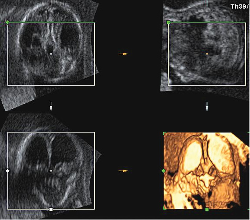
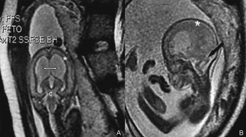
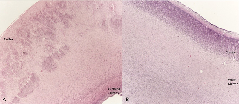
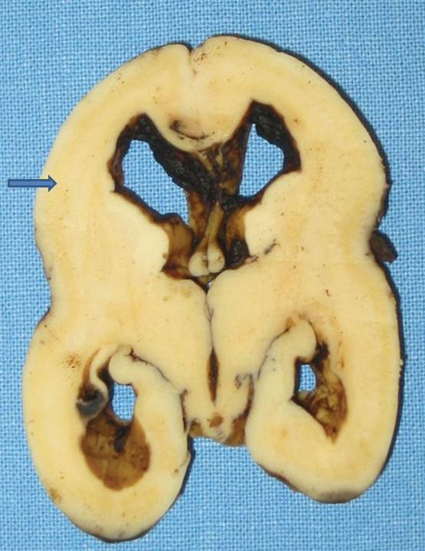
Lissencephaly is a genetic heterogeneous autosomal recessive disorder characterized by the classical triad: brain malformations, eye anomalies, and congenital muscular dystrophy. Prenatal diagnosis is feasible by demonstrating abnormal development of sulci and gyri. Magnetic resonance imaging (MRI) may enhance detection of developmental cortical disorders as well as ocular anomalies. We describe a case of early diagnosis of lissencephaly type 2 detected at the time of routine second trimester scan by three-dimensional ultrasound and fetal MRI. Gross pathology confirmed the accuracy of the prenatal diagnosis while histology showed the typical feature of cobblestone cortex. As the disease is associated with poor perinatal prognosis, early and accurate prenatal diagnosis is important for genetic counseling and antenatal care.
Resumo: Lissencefalia são doenças genéticas autossômicas recessivas heterogêneas caracterizadas pela tríade clássica: malformações do cérebro, anomalias oculares e distrofia muscular congênita. Diagnóstico pré-natal é factível pela demonstração do desenvolvimento anormal de sulcos e giros. Ressonância magnética (RM) melhora a detecção de distúrbios do desenvolvimento cortical, bem como as anomalias oculares. Descrevemos um caso de diagnóstico precoce de lisencefalia tipo 2 detectado no momento do ultrassom morfológico de segundo trimestre pela ultrassonografia tridimensional e RM fetal. A macroscopia confirmou a acurácia do diagnóstico pré-natal, enquanto que a microscopia mostrou a típica característica de córtex em cobblestone. Como a doença está associada à um pobre prognóstico perinatal, o precoce e acurado diagnóstico pré-natal é importante para o aconselhamento genético e seguimento da gestação.
Thieme Publicações Ltda Rio de Janeiro, Brazil.
Figures
References
-
- Walker W. Lissencephaly. Arch Neurol Psychiatry. 1942;48(1):13–29.
-
- Monteagudo A, Alayón A, Mayberry P. Walker-Warburg syndrome: case report and review of the literature. J Ultrasound Med. 2001;20(4):419–426. - PubMed
-
- Blin G, Rabbé A, Ansquer Y, Meghdiche S, Floch-Tudal C, Mandelbrot L. First-trimester ultrasound diagnosis in a recurrent case of Walker-Warburg syndrome. Ultrasound Obstet Gynecol. 2005;26(3):297–299. - PubMed
Publication types
MeSH terms
LinkOut - more resources
Full Text Sources
Other Literature Sources
Medical
Research Materials