Review
. 2016 May;271(1):114-26.
doi: 10.1111/imr.12398.

T-cell selection in the thymus: a spatial and temporal perspective

Affiliations
Review

T-cell selection in the thymus: a spatial and temporal perspective

Nadia Kurd et al. Immunol Rev. 2016 May.

Abstract

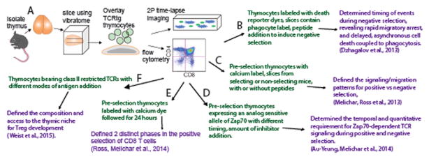
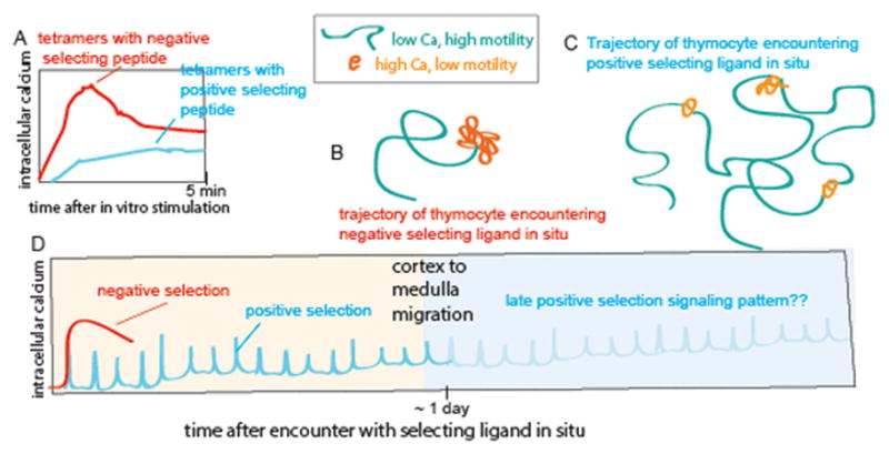
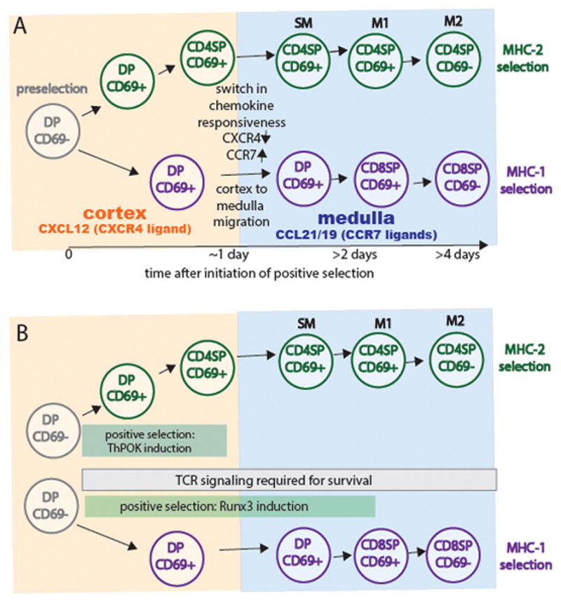
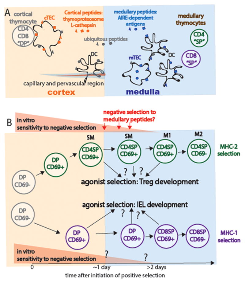
The ability of T cells to respond to a wide array of foreign antigens while avoiding reactivity to self is largely determined by cellular selection of developing T cells in the thymus. While a great deal is known about the cell types and molecules involved in T-cell selection in the thymus, our understanding of the spatial and temporal aspects of this process remain relatively poorly understood. Thymocytes are highly motile within the thymus and travel between specialized microenvironments at different phases of their development while interacting with distinct sets of self-peptides and peptide presenting cells. A knowledge of when, where, and how thymocytes encounter self-peptide MHC ligands at different stages of thymic development is key to understanding T-cell selection. In the past several years, our laboratory has investigated this topic using two-photon time-lapse microscopy to directly visualize thymocyte migration and signaling events, together with a living thymic slice preparation to provide a synchronized experimental model of T-cell selection in situ. Here, we discuss recent advances in our understanding of the temporal and spatial aspects of T-cell selection, highlighting our own work, and placing them in the context of work from other groups.

Keywords: T-cell antigen receptor; cell differentiation; chemokines; lineage commitment/specification; thymus.


Conflict of interest statement

The authors declare no conflict of interest.

Figures

Figure 1
Figure 1. Thymic tissue slices provide a robust and versatile system for the study of T cell selection
A) Thymocytes of defined TCR specificity from TCR transgenic mice are overlaid onto vibratome-cut slices of thymic tissue and cultured for up to three days. Thymocytes migrate to their normal location within the tissue according to chemokine gradients and undergo synchronized development, and can be examined using flow cytometry or 2-photon time-lapse imaging. B–F) Examples of variations of the thymic slice model (in green) and the relevant research advance (in purple). See text and references for details.
Figure 2
Figure 2. Temporal patterns of TCR signaling in vitro and in thymic slices
A) Temporal responses to stimulation of pre-selection thymocytes with MHC-tetramers loaded with low or high potency ligands. Adapted from (17). Stimulation with low potency peptide ligands in vitro leads to sustained low-level signaling. B, C) Schematic of representative trajectories of preselection thymocytes within thymic slices during encounters with negative (B) or positive (C) selecting ligands. Green indicates periods of low intracellular calcium and relatively rapid migration, where as orange indicates migratory pauses and elevated intracellular calcium levels. These schematics are based on data from 2-photon microscopy analysis of class I specific (OT1 or F5 TCR transgenic) thymocytes in thymic tissue slices. Using this system we have shown that negative selection correlates with a persistent increase in intracellular calcium, migratory arrest, and thymocyte death within 4–12 hours after the initiation of TCR signaling (10). In contrast, positive selection correlates with serial transient increases in intracellular calcium accompanied by migratory pauses interspersed with periods of rapid migration and low intracellular calcium (14). (D) The pattern of TCR signaling during the first 24 hours of positive selection (cyan) and negative selection (red) inferred from calcium signaling and motility changes in thymic slices. Thymocytes undergo a gradual increase in migratory speed and basal calcium levels throughout the first 24 hours of positive selection, while exhibiting progressively briefer transient signals. At around 24 hours thymocytes change their chemokine receptor expression and migrate from the cortex to the medulla (16). While it has been shown that TCR signaling is required late during positive selection (30, 31), the late signaling pattern has not yet been directly examined (indicated by faint portion of the curve). In addition, while it has also been reported that thymocytes bearing class II specific TCRs also undergo transient signals during positive selection (9), the signaling pattern associated with thymic positive selection on class II MHC has not yet been extensively examined.
Figure 3
Figure 3. Kinetics of chemokine receptor changes, intrathymic migration, co-receptor down-regulation during positive selection of class II versus class I restricted thymocytes
A) Preselection CD4+CD8+ DP thymocyte reside in the cortex and express CXCR4, the receptor for the cortical chemokine, CXCL12. More mature CD4+CD8− and CD4-CD8+ single positive (SP) thymocytes localize to the medulla and express CCR7, the receptor for the medullary chemokines CCL21/CCL19. Thymocytes undergoing selection via class II MHC first down-regulate CD8 and then change their chemokine receptor pattern around 24 hours after the initiation of positive selection. In contrast, although thymocytes undergoing selection via class I MHC also change their chemokine receptor expression around 24 hours, they do not down regulate CD4 until >1 day later. In the medulla, CD4SP thymocytes undergo a TCR independent maturation process (passing through stages termed semimature SM, M1, and M2) that correlate with down regulation of CD24 (HSA), upregulation of Qa2 and MHC class I, and the ability of thymocytes to die or proliferate upon TCR signaling (6, 34). CD8 SP thymocytes appear to progress through similar stages, although this has been less well studied. B) Thymocytes undergoing positive selection via class II MHC upregulate the CD4 defining transcription factor ThPOK and down-regulate CD8 after 24 hours of Zap70 dependent TCR signaling. In contrast, thymocytes undergoing positive selection via class I MHC require Zap70-dependent TCR signaling for an additional day or more in order to upregulate the CD8-defining transcription factor Runx3 and downregulate CD4. Both lineages continue to require TCR signaling to promote cell survival after lineage commitment and co-receptor downregulation (30, 66).
Figure 4
Figure 4. Distinct peptide and peptide presenting cells in the cortex versus medulla, and the timing of negative and agonist selection
Distinct peptide display and peptide presenting cells in the thymic cortex versus medulla. Cortical thymic epithelial cells (cTECs) express a unique proteasome subunit (the thymoproteasome) and lysosomal protease (L-cathepsin) that allows the generation of a unique peptide repertoire (orange) thought to promote positive selection (reviewed in (67)). In contrast, expression of the transcription factor Aire allows for the expression of tissue restricted antigens (blue) by medullary epithelial cells (mTECs). These tissue-restricted antigens can also be transferred to DCs, which are more prevalent within the medulla. Thus, thymocytes do not have access to tissue-restricted antigens until the positive-selection induced migration to the medulla. Thymocytes are exposed to ubiquitous self-peptides (gray) at all stages of development. Perivascular regions surrounding cortical capillaries have some medullary character, including the presence of the “medullary chemokine CCL21 and dendritic cells (36). B) Timing of negative and agonist selection. Thymocytes gradually lose their susceptibility to negative selection as they progress through progress from DP through CD4 SP stages “semi-mature” SM stage. While SM thymocytes undergo apoptosis upon strong TCR stimulation, M1 and M2 thymocytes become activated and proliferated (6, 34). The susceptibility of class I restricted thymocytes has been less well studied, but appears to follow a similar pattern. Thymocytes that encounter high affinity self-peptide ligands may undergo agonist selection rather than negative selection, giving rise to Tregs or CD8αα IEL.

References

    1. Klein L, Kyewski B, Allen PM, Hogquist KA. Positive and negative selection of the T cell repertoire: what thymocytes see (and don’t see) Nat Rev Immunol. 2014;14:377–391. - PMC - PubMed
    1. Love PE, Bhandoola A. Signal integration and crosstalk during thymocyte migration and emigration. Nat Rev Immunol. 2011;11:469–477. - PMC - PubMed
    1. Ladi E, Yin X, Chtanova T, Robey EA. Thymic microenvironments for T cell differentiation and selection. Nat Immunol. 2006;7:338–343. - PubMed
    1. Takahama Y. Journey through the thymus: stromal guides for T-cell development and selection. Nat Rev Immunol. 2006;6:127–135. - PubMed
    1. Starr TK, Jameson SC, Hogquist KA. Positive and negative selection of T cells. Annu Rev Immunol. 2003;21:139–176. - PubMed

Publication types