Review
. 2016 May;271(1):230-45.
doi: 10.1111/imr.12400.

Peyer's patches: organizing B-cell responses at the intestinal frontier

Affiliations
Review

Peyer's patches: organizing B-cell responses at the intestinal frontier

Andrea Reboldi et al. Immunol Rev. 2016 May.

Abstract

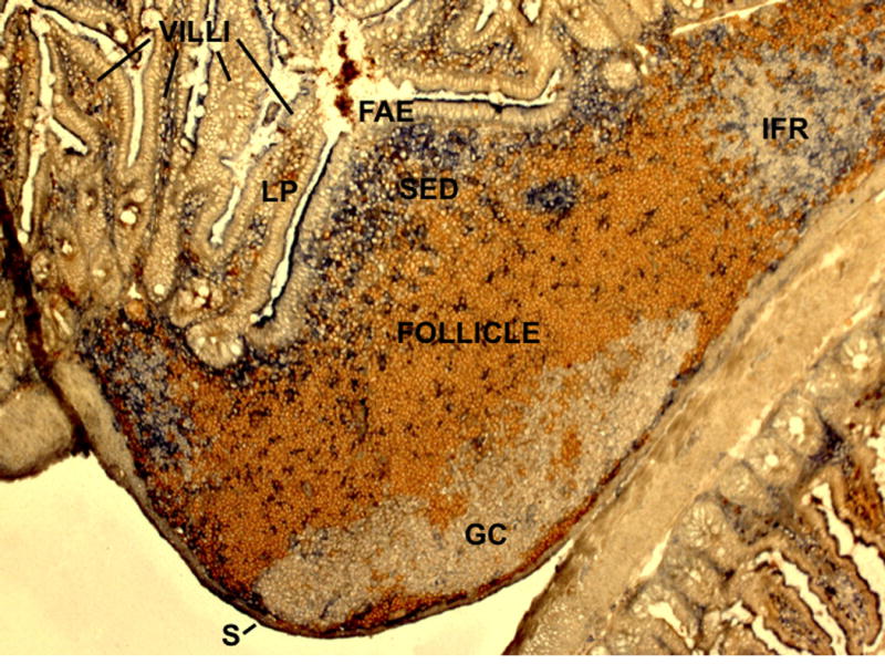
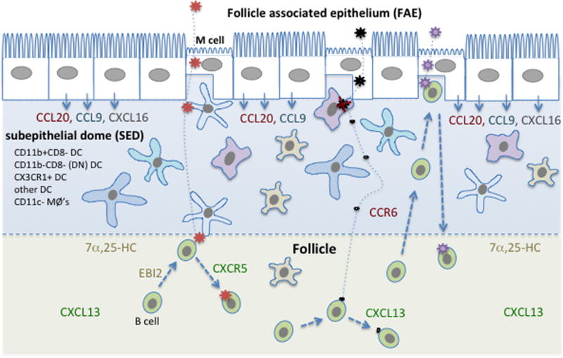
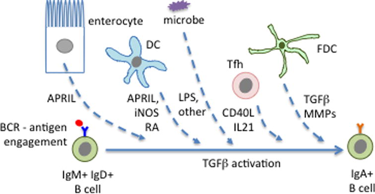
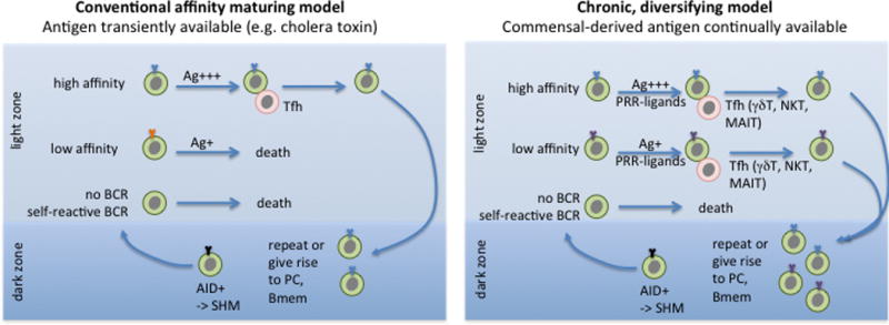
Secondary lymphoid tissues share the important function of bringing together antigens and rare antigen-specific lymphocytes to foster induction of adaptive immune responses. Peyer's patches (PPs) are unique compared to other secondary lymphoid tissues in their continual exposure to an enormous diversity of microbiome- and food-derived antigens and in the types of pathogens they encounter. Antigens are delivered to PPs by specialized microfold (M) epithelial cells and they may be captured and presented by resident dendritic cells (DCs). In accord with their state of chronic microbial antigen exposure, PPs exhibit continual germinal center (GC) activity. These GCs not only contribute to the generation of B cells and plasma cells producing somatically mutated gut antigen-specific IgA antibodies but have also been suggested to support non-specific antigen diversification of the B-cell repertoire. Here, we review current understanding of how PPs foster B-cell encounters with antigen, how they favor isotype switching to the secretory IgA isotype, and how their GC responses may uniquely contribute to mucosal immunity.

Keywords: B cells; antibodies; cell trafficking; dendritic cells; mucosa; repertoire development.


Conflict of interest statement

The authors declare they have no financial conflict of interest

Figures

Figure 1
Figure 1. Cross-sectional view of mouse Peyer’s patch showing main anatomical compartments
Stained to detect naïve B cells (IgD, brown) and dendritic cells (CD11c, blue). FAE, follicle associated epithelium; SED, subepithelial dome; GC, germinal center; IFR, interfollicular region (T cell zone); LP, lamina propria; S, serosa. During embedding of the tissue for preparation of the 7 μm frozen sections the PP became juxtaposed to another loop of small intestine (bottom right of the image).
Figure 2
Figure 2. Diagram highlighting cellular complexity of the PP subepithelial dome (SED) and showing several possible mechanisms of B cell antigen exposure
Major myeloid cell types present in the SED are listed and represented by different cell shapes. Distribution of major chemoattractant factors is indicated. Stromal cells, which include the cells that produce the CXCR5 ligand CXCL13 and the EBI2 ligand, 7α,25-HC, are not shown. Three example antigens are shown entering the SED after M cell-mediated transcytosis. The red antigen then travels via DC to reach the interface with the follicle. B cell movement to this interface is favored by EBI2 responding to 7α,25-HC. The black antigen is taken up by a macrophage and largely degraded, but some small fragments travel away passively and reach follicular B cells. The purple antigen is encountered directly by a B cell that is suggested to have accessed the SED and M cell pocket in a CCR6-CCL20 dependent manner, likely a recently activated or memory B cell.
Figure 3
Figure 3. Factors promoting IgA switching in PPs
The highlighted factors are known to be produced by the indicated cell types but in several cases it has not yet been established which is the key cellular source of the factor and whether the action on the B cell is direct or indirect. Microbe-derived ligands for pattern recognition receptors, such as LPS, may also promote switching. See text for further details.
Figure 4
Figure 4. Two models of GC function in PPs
In the conventional model, GC B cells that acquire a high affinity BCR through AID-dependent SHM in the dark zone take up more of the transiently available antigen (Ag) than low affinity cells while in the light zone and win-out in receiving T cell help, leading to their selective survival and continuation on in the GC reaction or emergence as high affinity memory B cells (Bmem) or plasma cells (PC). In the diversifying model, there is continually available antigen, and sufficient T cell help (possibly of a range of types) to support the survival and differentiation of low and high affinity cells. Commensal-derived signals acting via pattern-recognition receptors (PRRs) likely cooperate with T cell-derived signals to support the chronic GC responses.

References

    1. Brandtzaeg P. Role of secretory antibodies in the defence against infections. Int J Med Microbiol. 2003;293:3–15. - PubMed
    1. Blutt SE, Conner ME. The gastrointestinal frontier: IgA and viruses. Front Immunol. 2013;4:402. - PMC - PubMed
    1. Macpherson AJ, Koller Y, McCoy KD. The bilateral responsiveness between intestinal microbes and IgA. Trends Immunol. 2015;36:460–470. - PubMed
    1. Fagarasan S, Kawamoto S, Kanagawa O, Suzuki K. Adaptive immune regulation in the gut: T cell-dependent and T cell-independent IgA synthesis. Annu Rev Immunol. 2010;28:243–273. - PubMed
    1. Macpherson AJ, Geuking MB, Slack E, Hapfelmeier S, McCoy KD. The habitat, double life, citizenship, and forgetfulness of IgA. Immulogical Reviews. 2011;245:132–146. - PubMed

Publication types

Substances